
Deposition Date
2004-12-30
Release Date
2005-05-24
Last Version Date
2024-10-30
Entry Detail
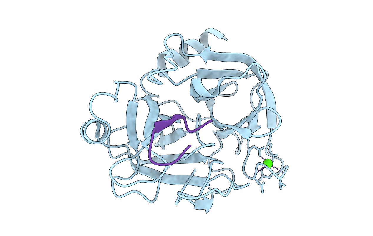
PDB ID:
1YF4
Keywords:
Title:
Crystal Structure of trypsin-vasopressin complex
Biological Source:
Source Organism(s):
Sus scrofa (Taxon ID: 9823)
Method Details:
Experimental Method:
Resolution:
1.98 Å
R-Value Free:
0.22
R-Value Work:
0.18
R-Value Observed:
0.18
Space Group:
P 21 21 21
