1YE6 image
Deposition Date 2004-12-28
Release Date 2005-02-01
Last Version Date 2023-08-23
Entry Detail
PDB ID:
1YE6
Keywords:
Title:
Crystal structure of the Lys-274 to Arg mutant of Candida tenuis xylose reductase (AKR2B5) bound to NADP+
Biological Source:
Source Organism:
Candida tenuis (Taxon ID: 45596)
Method Details:
Experimental Method:
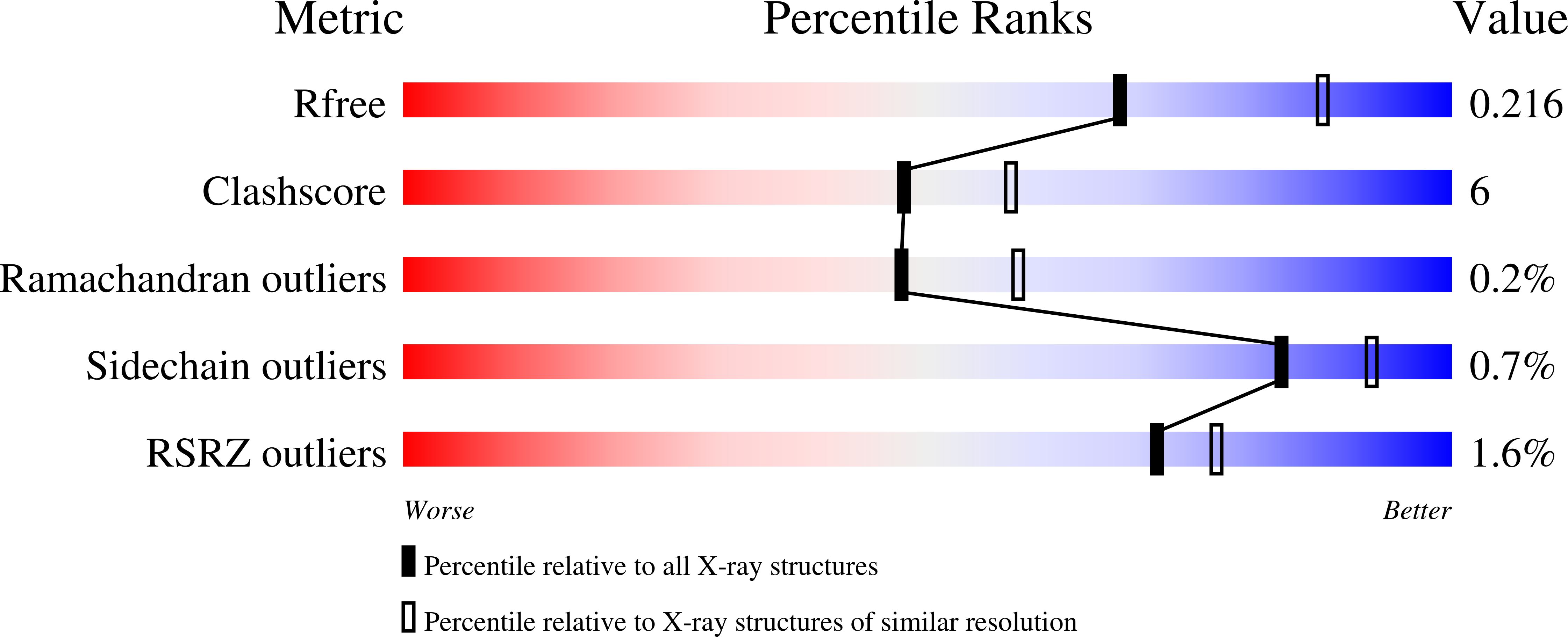
Resolution:
2.30 Å
R-Value Free:
0.22
R-Value Work:
0.19
R-Value Observed:
0.19
Space Group:
C 1 2 1
Macromolecular Entities
Polymer Type:polypeptide(L)
Molecule:NAD(P)H-dependent D-xylose reductase
Gene (Uniprot):XYL1
Mutations:K274R
Chain IDs:A, B, C, D
Chain Length:322
Number of Molecules:4
Biological Source:Candida tenuis
Primary Citation
Fine tuning of coenzyme specificity in family 2 aldo-keto reductases revealed by crystal structures of the Lys-274-->Arg mutant of Candida tenuis xylose reductase (AKR2B5) bound to NAD(+) and NADP(+).
Febs Lett. 579 763 767 (2005)
PMID: 15670843 DOI: 10.1016/j.febslet.2004.12.063

Abstact

Aldo-keto reductases of family 2 employ single site replacement Lys-->Arg to switch their cosubstrate preference from NADPH to NADH. X-ray crystal structures of Lys-274-->Arg mutant of Candida tenuis xylose reductase (AKR2B5) bound to NAD+ and NADP+ were determined at a resolution of 2.4 and 2.3A, respectively. Due to steric conflicts in the NADP+-bound form, the arginine side chain must rotate away from the position of the original lysine side chain, thereby disrupting a network of direct and water-mediated interactions between Glu-227, Lys-274 and the cofactor 2'-phosphate and 3'-hydroxy groups. Because anchoring contacts of its Glu-227 are lost, the coenzyme-enfolding loop that becomes ordered upon binding of NAD(P)+ in the wild-type remains partly disordered in the NADP+-bound mutant. The results delineate a catalytic reaction profile for the mutant in comparison to wild-type.

Legend

Protein

Chemical

Disease

Primary Citation of related structures