1YDX image
Deposition Date 2004-12-27
Release Date 2005-08-23
Last Version Date 2024-11-20
Entry Detail
PDB ID:
1YDX
Title:
Crystal structure of Type-I restriction-modification system S subunit from M. genitalium
Biological Source:
Source Organism:
Host Organism:
Method Details:
Experimental Method:
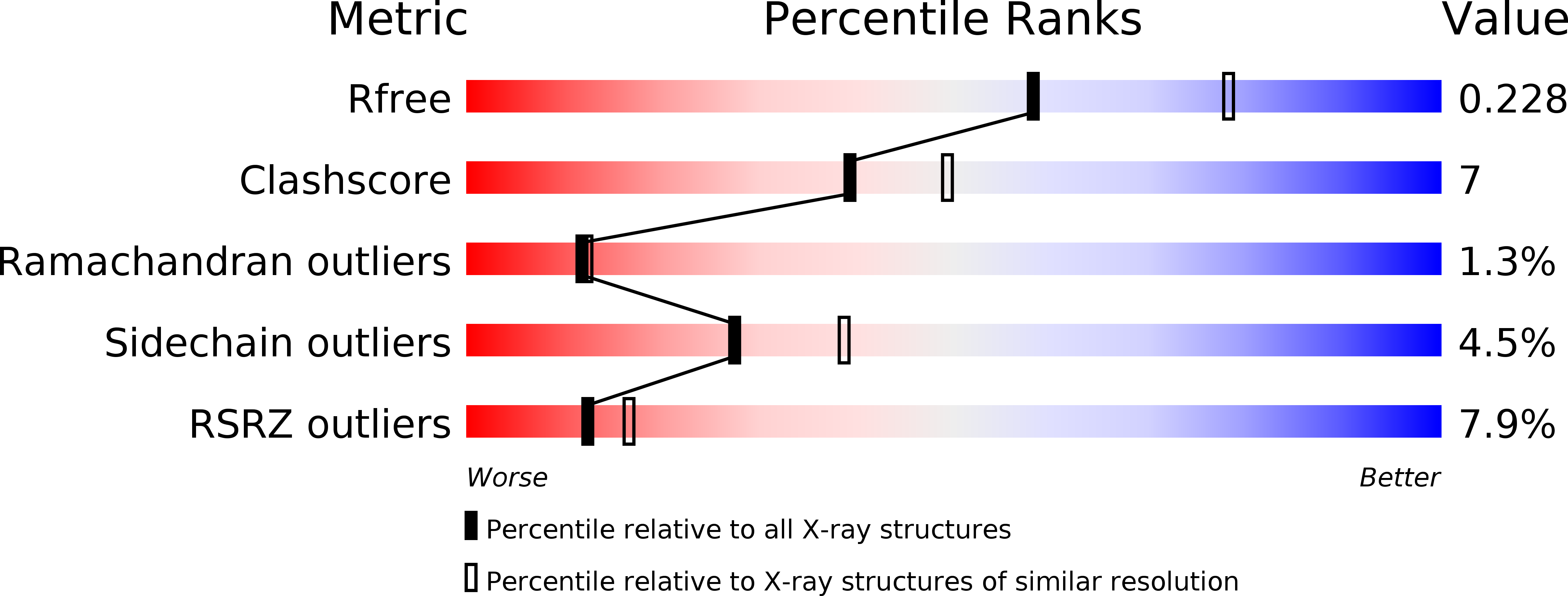
Resolution:
2.30 Å
R-Value Free:
0.23
R-Value Work:
0.19
R-Value Observed:
0.19
Space Group:
P 31 2 1
Macromolecular Entities
Structures with similar UniProt ID
Protein Blast
Polymer Type:polypeptide(L)
Molecule:type I restriction enzyme specificity protein MG438
Gene (Uniprot):MG438
Chain IDs:A
Chain Length:406
Number of Molecules:1
Biological Source:Mycoplasma genitalium
Modified Residue
Compound ID Chain ID Parent Comp ID Details 2D Image
MSE A MET SELENOMETHIONINE
Ligand Molecules
Primary Citation
Crystal Structure of a Putative Type I Restriction-Modification S Subunit from Mycoplasma genitalium
J.Mol.Biol. 351 749 762 (2005)
PMID: 16038930 DOI: 10.1016/j.jmb.2005.06.050

Abstact

The crystal structure of the eubacteria Mycoplasma genitalium ORF MG438 polypeptide, determined by multiple anomalous dispersion and refined at 2.3 A resolution, reveals the organization of S subunits from the Type I restriction and modification system. The structure consists of two globular domains, with about 150 residues each, separated by a pair of 40 residue long antiparallel alpha-helices. The globular domains correspond to the variable target recognition domains (TRDs), as previously defined for S subunits on sequence analysis, while the two helices correspond to the central (CR1) and C-terminal (CR2) conserved regions, respectively. The structure of the MG438 subunit presents an overall cyclic topology with an intramolecular 2-fold axis that superimposes the N and the C-half parts, each half containing a globular domain and a conserved helix. TRDs are found to be structurally related with the small domain of the Type II N6-adenine DNA MTase TaqI. These relationships together with the structural peculiarities of MG438, in particular the presence of the intramolecular quasi-symmetry, allow the proposal of a model for S subunits recognition of their DNA targets in agreement with previous experimental results. In the crystal, two subunits of MG438 related by a crystallographic 2-fold axis present a large contact area mainly involving the symmetric interactions of a cluster of exposed hydrophobic residues. Comparison with the recently reported structure of an S subunit from the archaea Methanococcus jannaschii highlights the structural features preserved despite a sequence identity below 20%, but also reveals important differences in the globular domains and in their disposition with respect to the conserved regions.

Legend

Protein

Chemical

Disease

Primary Citation of related structures
Feedback Form
Name
Email
Institute
Feedback