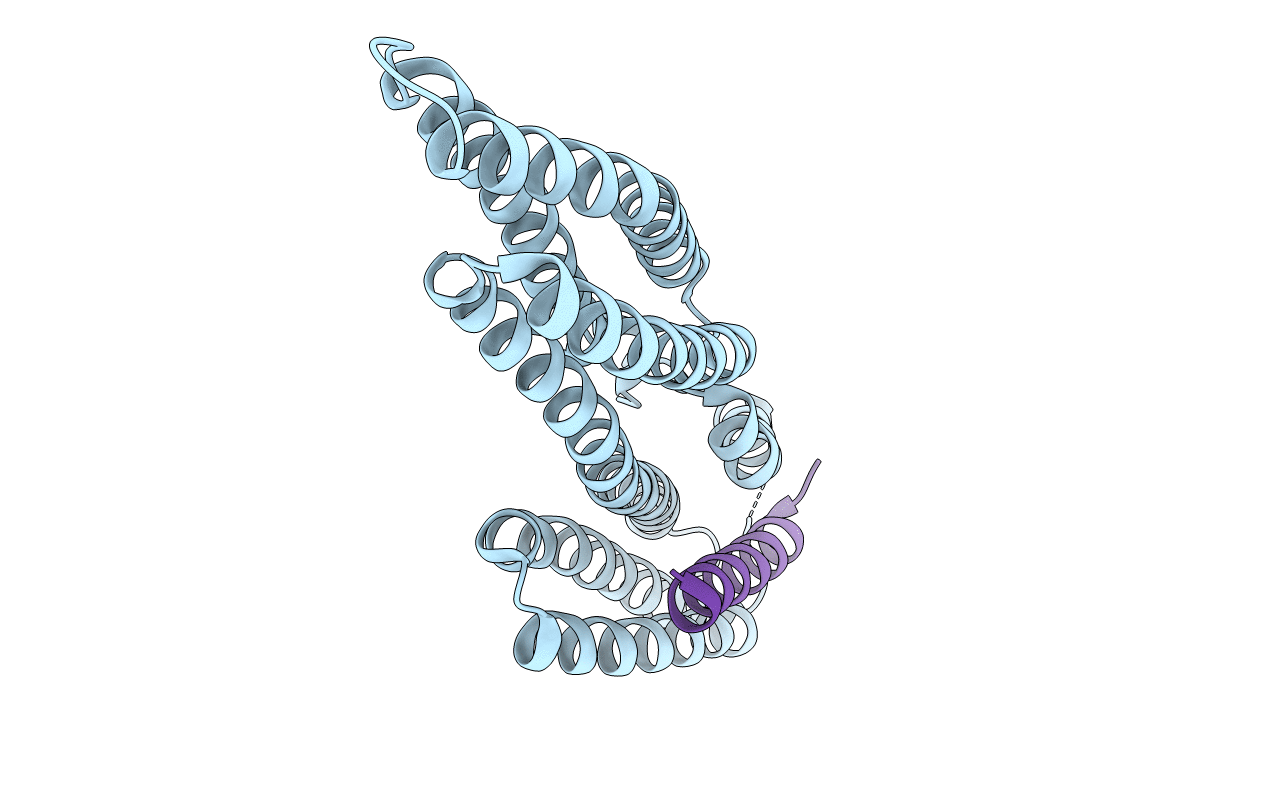
1YDI image
Deposition Date 2004-12-23
Release Date 2005-07-19
Last Version Date 2024-02-14
Entry Detail
PDB ID:
1YDI
Title:
Human Vinculin Head Domain (VH1, 1-258) in Complex with Human Alpha-Actinin's Vinculin-Binding Site (Residues 731-760)
Biological Source:
Source Organism:
Homo sapiens (Taxon ID: 9606)
Host Organism:
Method Details:
Experimental Method:
Resolution:
1.80 Å
R-Value Free:
0.23
R-Value Work:
0.17
R-Value Observed:
0.18
Space Group:
P 1 21 1
Macromolecular Entities
Structures with similar UniProt ID
Protein Blast
Polymer Type:polypeptide(L)
Molecule:vinculin isoform VCL
Chain IDs:A
Chain Length:263
Number of Molecules:1
Biological Source:Homo sapiens
Structures with similar UniProt ID
Protein Blast
Polymer Type:polypeptide(L)
Molecule:Alpha-actinin 4
Gene (Uniprot):ACTN4
Chain IDs:B
Chain Length:24
Number of Molecules:1
Biological Source:Homo sapiens
Primary Citation
Structural Dynamics of {alpha}-Actinin-Vinculin Interactions.
Mol.Cell.Biol. 14 6112 6122 (2005)
PMID: 15988023 DOI: 10.1128/MCB.25.14.6112-6122.2005

Abstact

Alpha-actinin and vinculin orchestrate reorganization of the actin cytoskeleton following the formation of adhesion junctions. alpha-Actinin interacts with vinculin through the binding of an alpha-helix (alphaVBS) present within the R4 spectrin repeat of its central rod domain to vinculin's N-terminal seven-helical bundle domain (Vh1). The Vh1:alphaVBS structure suggests that alphaVBS first unravels from its buried location in the triple-helical R4 repeat to allow it to bind to vinculin. alphaVBS binding then induces novel conformational changes in the N-terminal helical bundle of Vh1, which disrupt its intramolecular association with vinculin's tail domain and which differ from the alterations in Vh1 provoked by the binding of talin. Surprisingly, alphaVBS binds to Vh1 in an inverted orientation compared to the binding of talin's VBSs to vinculin. Importantly, the binding of alphaVBS and talin's VBSs to vinculin's Vh1 domain appear to also trigger distinct conformational changes in full-length vinculin, opening up distant regions that are buried in the inactive molecule. The data suggest a model where vinculin's Vh1 domain acts as a molecular switch that undergoes distinct structural changes provoked by talin and alpha-actinin binding in focal adhesions versus adherens junctions, respectively.

Legend

Protein

Chemical

Disease

Primary Citation of related structures