1YCH image
Deposition Date 2004-12-22
Release Date 2005-04-19
Last Version Date 2024-02-14
Entry Detail
PDB ID:
1YCH
Keywords:
Title:
X-ray Crystal Structures of Moorella thermoacetica FprA. Novel Diiron Site Structure and Mechanistic Insights into a Scavenging Nitric Oxide Reductase
Biological Source:
Source Organism:
Host Organism:
Method Details:
Experimental Method:
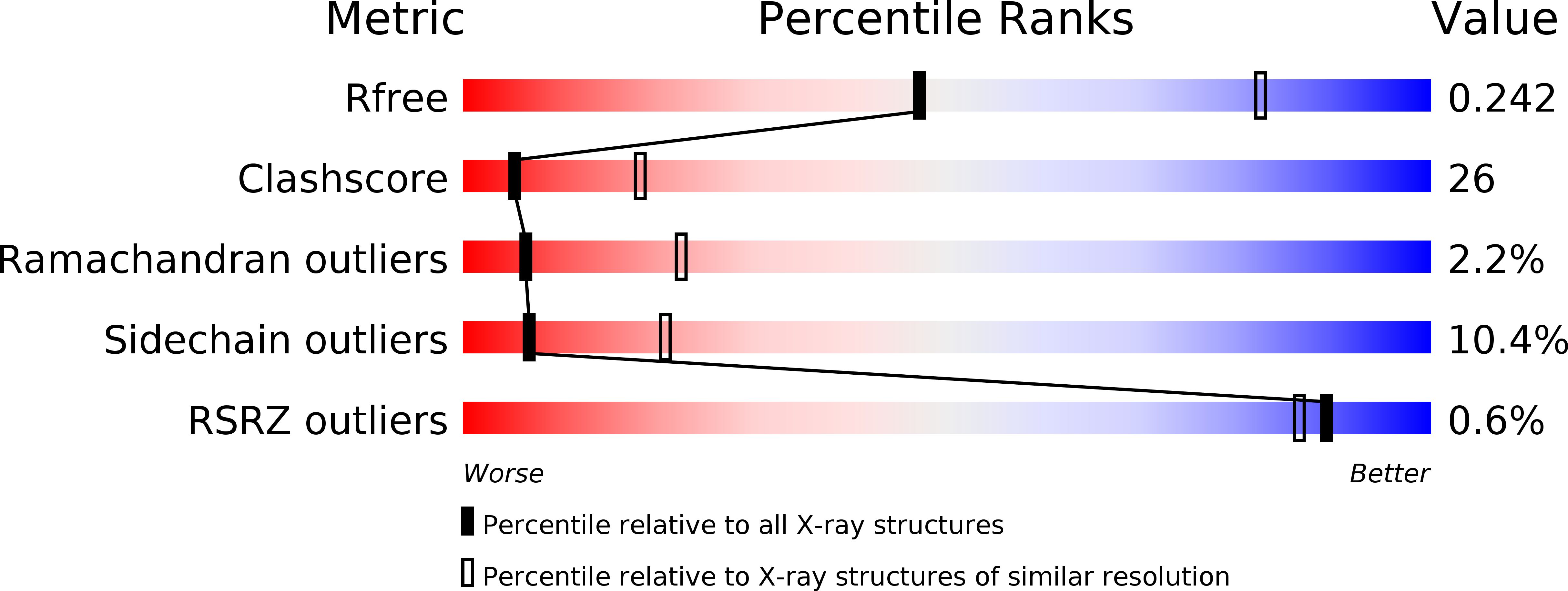
Resolution:
2.80 Å
R-Value Free:
0.24
R-Value Work:
0.22
R-Value Observed:
0.22
Space Group:
P 43 21 2
Macromolecular Entities
Structures with similar UniProt ID
Protein Blast
Polymer Type:polypeptide(L)
Molecule:Nitric oxide reductase
Gene (Uniprot):fprA
Chain IDs:A, B, C, D
Chain Length:398
Number of Molecules:4
Biological Source:Moorella thermoacetica
Primary Citation
X-ray crystal structures of Moorella thermoacetica FprA. Novel diiron site structure and mechanistic insights into a scavenging nitric oxide reductase.
Biochemistry 44 6492 6501 (2005)
PMID: 15850383 DOI: 10.1021/bi0473049

Abstact

Several members of a widespread class of bacterial and archaeal metalloflavoproteins, called FprA, likely function as scavenging nitric oxide reductases (S-NORs). However, the only published X-ray crystal structure of an FprA is for a protein characterized as a rubredoxin:dioxygen oxidoreductase (ROO) from Desulfovibrio gigas. Therefore, the crystal structure of Moorella thermoacetica FprA, which has been established to function as an S-NOR, was solved in three different states: as isolated, reduced, and reduced, NO-reacted. As is the case for D. gigas ROO, the M. thermoacetica FprA contains a solvent-bridged non-heme, non-sulfur diiron site with five-coordinate iron centers bridged by an aspartate, and terminal glutamate, aspartate, and histidine ligands. However, the M. thermoacetica FprA diiron site showed four His ligands, two to each iron, in all three states, whereas the D. gigas ROO diiron site was reported to contain only three His ligands, even though the fourth His residue is conserved. The Fe1-Fe2 distance within the diiron site of M. thermoacetica FprA remained at 3.2-3.4 A with little or no movement of the protein ligands in the three different states and with conservation of the two proximal open coordination sites. Molecular modeling indicated that each open coordination site can accommodate an end-on NO. This relatively rigid and symmetrical diiron site structure is consistent with formation of a diferrous dinitrosyl as the committed catalytic intermediate leading to formation of N(2)O. These results provide new insight into the structural features that fine-tune biological non-heme diiron sites for dioxygen activation vs nitric oxide reduction.

Legend

Protein

Chemical

Disease

Primary Citation of related structures