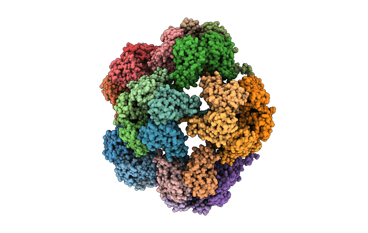
1YC6 image
Deposition Date 2004-12-22
Release Date 2005-01-18
Last Version Date 2023-08-23
Entry Detail
PDB ID:
1YC6
Keywords:
Title:
Crystallographic Structure of the T=1 Particle of Brome Mosaic Virus
Biological Source:
Source Organism:
Method Details:
Experimental Method:
Resolution:
2.90 Å
R-Value Free:
0.23
R-Value Work:
0.21
R-Value Observed:
0.22
Space Group:
P 43 2 2
Macromolecular Entities
Polymer Type:polypeptide(L)
Molecule:Coat protein
Gene (Uniprot):ORF3b
Chain IDs:A, B, C, D, E, F, G, H, I, J, K, L, M, N, O, P, Q, R, S, T, U, V, W, X, Y, Z, AA (auth: 1), BA (auth: 2), CA (auth: 3), DA (auth: 4)
Chain Length:154
Number of Molecules:30
Biological Source:Brome mosaic virus
Primary Citation
Crystallographic structure of the T=1 particle of brome mosaic virus.
J.Mol.Biol. 346 815 831 (2005)
PMID: 15713465 DOI: 10.1016/j.jmb.2004.12.015

Abstact

T=1 icosahedral particles of amino terminally truncated brome mosaic virus (BMV) protein were created by treatment of the wild-type T=3 virus with 1M CaCl2 and crystallized from sodium malonate. Diffraction data were collected from frozen crystals to beyond 2.9 A resolution and the structure determined by molecular replacement and phase extension. The particles are composed of pentameric capsomeres from the wild-type virions which have reoriented with respect to the original particle pentameric axes by rotations of 37 degrees , and formed tenuous interactions with one another, principally through conformationally altered C-terminal polypeptides. Otherwise, the pentamers are virtually superimposable upon those of the original T=3 BMV particles. The T=1 particles, in the crystals, are not perfect icosahedra, but deviate slightly from exact symmetry, possibly due to packing interactions. This suggests that the T=1 particles are deformable, which is consistent with the loose arrangement of pentamers and latticework of holes that penetrate the surface. Atomic force microscopy showed that the T=3 to T=1 transition could occur by shedding of hexameric capsomeres and restructuring of remaining pentamers accompanied by direct condensation. Knowledge of the structures of the BMV wild-type and T=1 particles now permit us to propose a tentative model for that process. A comparison of the BMV T=1 particles was made with the reassembled T=1 particles produced from the coat protein of trypsin treated alfalfa mosaic virus (AlMV), another bromovirus. There is little resemblance between the two particles. The BMV particle, with a maximum diameter of 195 A, is made from distinctive pentameric capsomeres with large holes along the 3-fold axis, while the AlMV particle, of approximate maximum diameter 220 A, has subunits closely packed around the 3-fold axis, large holes along the 5-fold axis, and few contacts within pentamers. In both particles crucial linkages are made about icosahedral dyads.

Legend

Protein

Chemical

Disease

Primary Citation of related structures