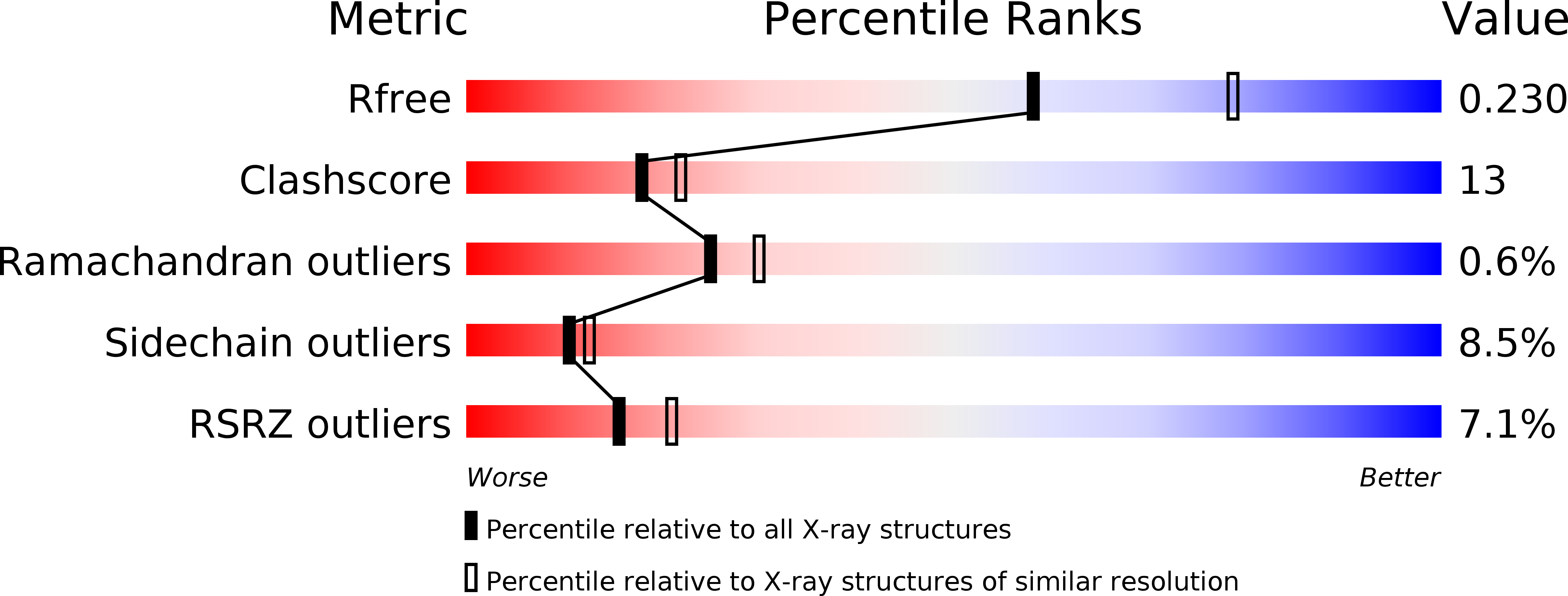
1YBT image
Deposition Date 2004-12-21
Release Date 2005-02-15
Last Version Date 2024-11-13
Entry Detail
PDB ID:
1YBT
Keywords:
Title:
MYCOBACTERIUM TUBERCULOSIS ADENYLYL CYCLASE, RV1900C CHD
Biological Source:
Source Organism:
Host Organism:
Method Details:
Experimental Method:
Resolution:
2.31 Å
R-Value Free:
0.23
R-Value Work:
0.19
R-Value Observed:
0.19
Space Group:
P 1 2 1
Macromolecular Entities
Structures with similar UniProt ID
Protein Blast
Polymer Type:polypeptide(L)
Molecule:hydrolase, alpha/beta hydrolase fold family
Gene (Uniprot):lipJ
Chain IDs:A, B, C, D
Chain Length:184
Number of Molecules:4
Biological Source:Mycobacterium tuberculosis
Modified Residue
Compound ID Chain ID Parent Comp ID Details 2D Image
MSE A MET SELENOMETHIONINE
Primary Citation
Origin of asymmetry in adenylyl cyclases: structures of Mycobacterium tuberculosis Rv1900c.
Embo J. 24 663 673 (2005)
PMID: 15678099 DOI: 10.1038/sj.emboj.7600573

Abstact

Rv1900c, a Mycobacterium tuberculosis adenylyl cyclase, is composed of an N-terminal alpha/beta-hydrolase domain and a C-terminal cyclase homology domain. It has an unusual 7% guanylyl cyclase side-activity. A canonical substrate-defining lysine and a catalytic asparagine indispensable for mammalian adenylyl cyclase activity correspond to N342 and H402 in Rv1900c. Mutagenic analysis indicates that these residues are dispensable for activity of Rv1900c. Structures of the cyclase homology domain, solved to 2.4 A both with and without an ATP analog, form isologous, but asymmetric homodimers. The noncanonical N342 and H402 do not interact with the substrate. Subunits of the unliganded open dimer move substantially upon binding substrate, forming a closed dimer similar to the mammalian cyclase heterodimers, in which one interfacial active site is occupied and the quasi-dyad-related active site is occluded. This asymmetry indicates that both active sites cannot simultaneously be catalytically active. Such a mechanism of half-of-sites-reactivity suggests that mammalian heterodimeric adenylyl cyclases may have evolved from gene duplication of a primitive prokaryote-type cyclase, followed by loss of function in one active site.

Legend

Protein

Chemical

Disease

Primary Citation of related structures
Feedback Form
Name
Email
Institute
Feedback