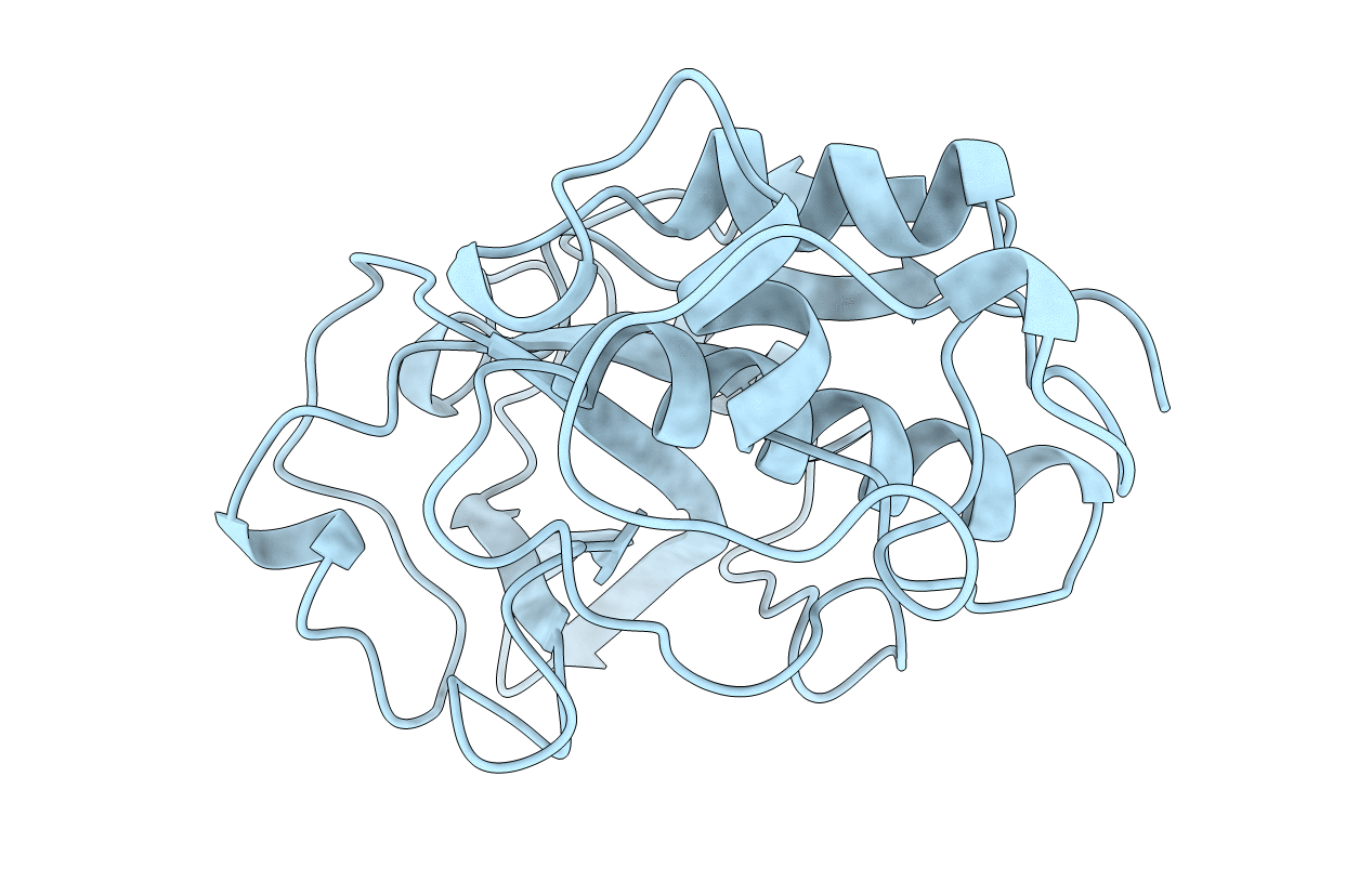
1YAL image
Deposition Date 1996-06-20
Release Date 1996-12-23
Last Version Date 2023-08-09
Entry Detail
PDB ID:
1YAL
Keywords:
Title:
CARICA PAPAYA CHYMOPAPAIN AT 1.7 ANGSTROMS RESOLUTION
Biological Source:
Source Organism:
Carica papaya (Taxon ID: 3649)
Method Details:
Experimental Method:
Resolution:
1.70 Å
R-Value Work:
0.19
R-Value Observed:
0.19
Space Group:
C 1 2 1
Macromolecular Entities
Structures with similar UniProt ID
Protein Blast
Polymer Type:polypeptide(L)
Molecule:CHYMOPAPAIN
Chain IDs:A
Chain Length:218
Number of Molecules:1
Biological Source:Carica papaya
Modified Residue
Compound ID Chain ID Parent Comp ID Details 2D Image
SCH A CYS S-METHYL-THIO-CYSTEINE
Primary Citation
Structure of chymopapain at 1.7 A resolution.
Biochemistry 35 16292 16298 (1996)
PMID: 8973203 DOI: 10.1021/bi961491w

Abstact

The X-ray structure of chymopapain, a cysteine proteinase isolated from the latex of the fruits of Carica papaya L., has been determined by molecular replacement methods and refined to a conventional R factor of 0.19 for all observed reflections in the range from 9.5 to 1.7 A resolution. The crystals used in this study contained a unique molecular species of chymopapain with two moles of thiomethyl attached to the two free cysteines per mole of enzyme. A comparison is made with the other known papaya proteinase X-ray structures: papain, caricain, and glycyl endopeptidase. Their backbone conformations are extremely similar except for two loop regions. Both regions are located at the surface of the protein and far away of the active site cleft. In each X-ray structure the same water network was found at the interface between the two domains of the enzyme. A close examination of the active site groove showed that the specificity restrictions dictated by the S2 subsite did not differ significantly among the four proteinases.

Legend

Protein

Chemical

Disease

Primary Citation of related structures