Deposition Date
2004-12-17
Release Date
2005-06-07
Last Version Date
2023-10-25
Entry Detail
PDB ID:
1YAB
Keywords:
Title:
Structure of T. maritima FliN flagellar rotor protein
Biological Source:
Source Organism(s):
Thermotoga maritima (Taxon ID: 243274)
Expression System(s):
Method Details:
Experimental Method:
Resolution:
3.40 Å
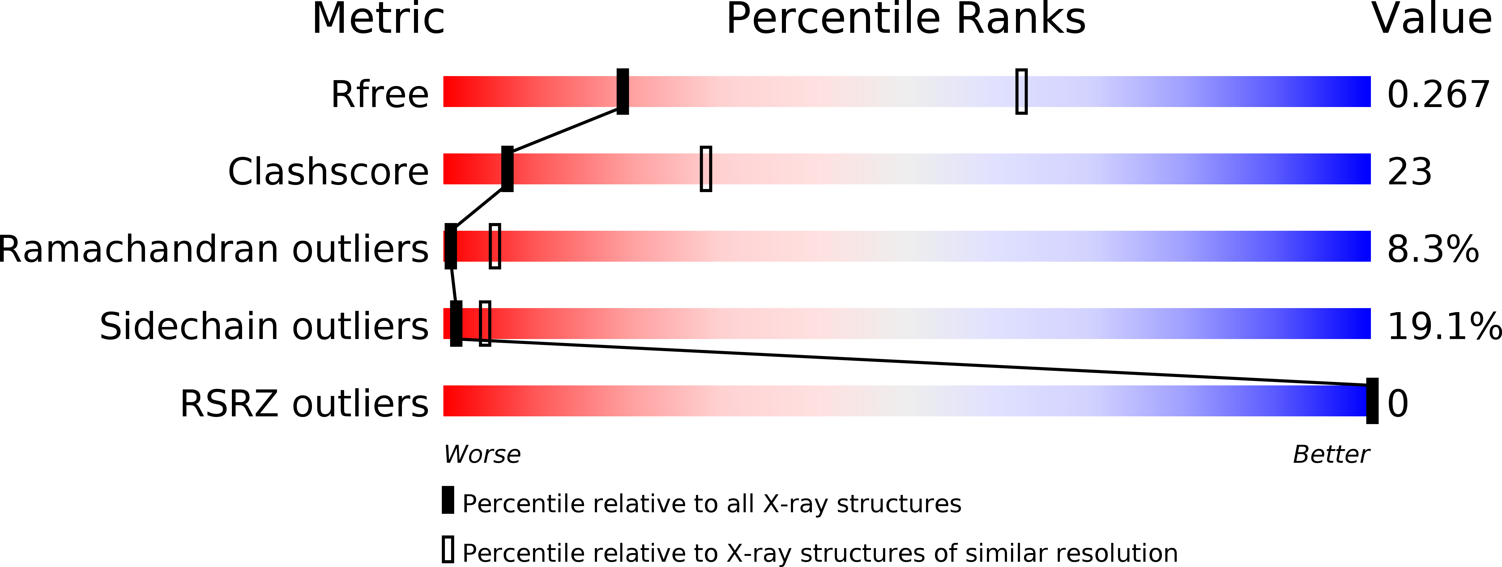
R-Value Free:
0.28
R-Value Work:
0.22
R-Value Observed:
0.22
Space Group:
P 31 2 1