1Y9L image
Deposition Date 2004-12-15
Release Date 2005-04-12
Last Version Date 2024-11-20
Entry Detail
PDB ID:
1Y9L
Title:
The X-ray structure of an secretion system protein
Biological Source:
Source Organism:
Method Details:
Experimental Method:
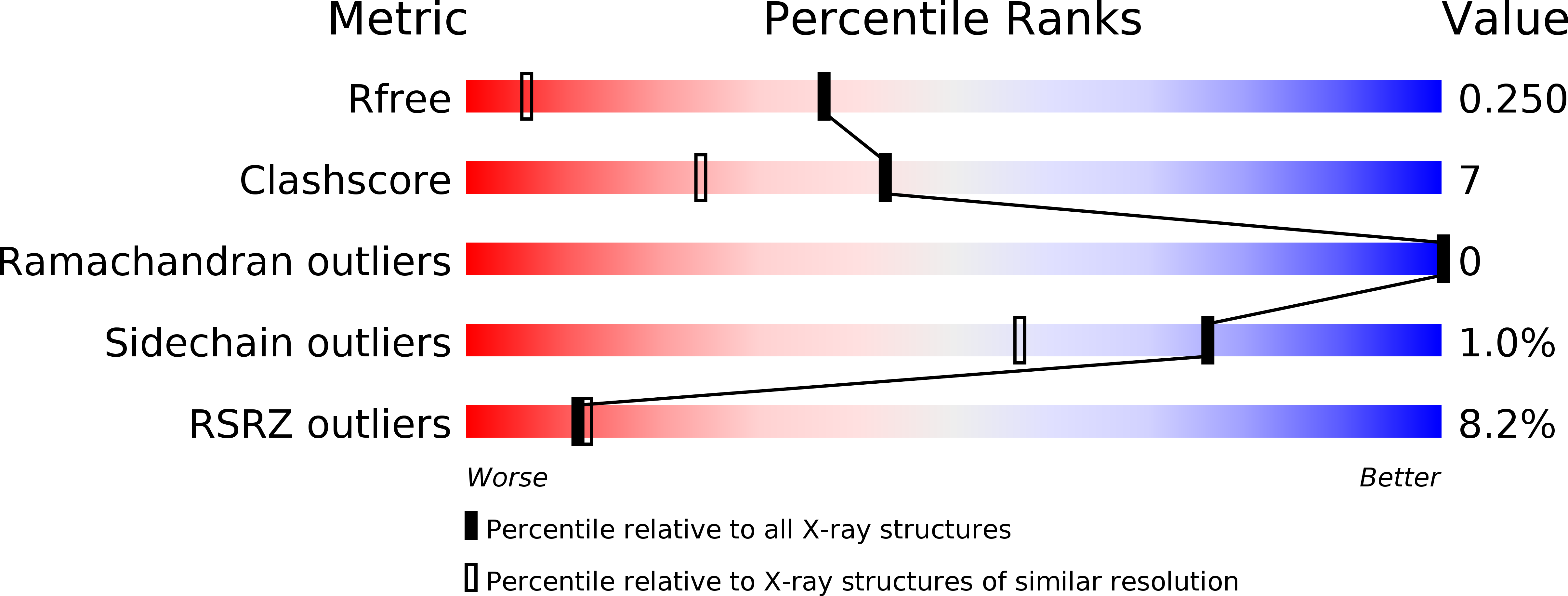
Resolution:
1.50 Å
R-Value Free:
0.25
R-Value Work:
0.22
R-Value Observed:
0.22
Space Group:
C 1 2 1
Macromolecular Entities
Polymer Type:polypeptide(L)
Molecule:Lipoprotein mxiM
Gene (Uniprot):mxiM
Chain IDs:A
Chain Length:115
Number of Molecules:1
Biological Source:Shigella flexneri
Primary Citation
Structure and biochemical analysis of a secretin pilot protein.
Embo J. 24 1111 1121 (2005)
PMID: 15775974 DOI: 10.1038/sj.emboj.7600610

Abstact

The ability to translocate virulence proteins into host cells through a type III secretion apparatus (TTSS) is a hallmark of several Gram-negative pathogens including Shigella, Salmonella, Yersinia, Pseudomonas, and enteropathogenic Escherichia coli. In common with other types of bacterial secretion apparatus, the assembly of the TTSS complex requires the preceding formation of its integral outer membrane secretin ring component. We have determined at 1.5 A the structure of MxiM28-142, the Shigella pilot protein that is essential for the assembly and membrane association of the Shigella secretin, MxiD. This represents the first atomic structure of a secretin pilot protein from the several bacterial secretion systems containing an orthologous secretin component. A deep hydrophobic cavity is observed in the novel 'cracked barrel' structure of MxiM, providing a specific binding domain for the acyl chains of bacterial lipids, a proposal that is supported by our various lipid/MxiM complex structures. Isothermal titration analysis shows that the C-terminal domain of the secretin, MxiD525-570, hinders lipid binding to MxiM.

Legend

Protein

Chemical

Disease

Primary Citation of related structures