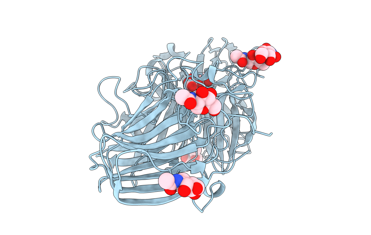
1Y9G image
Entry Detail
PDB ID:
1Y9G
Keywords:
Title:
Crystal structure of exo-inulinase from Aspergillus awamori complexed with fructose
Biological Source:
Source Organism:
PDB Version:
Deposition Date:
2004-12-15
Release Date:
2004-12-21
Method Details:
Experimental Method:
Resolution:
1.87 Å
R-Value Free:
0.19
R-Value Work:
0.14
R-Value Observed:
0.15
Space Group:
P 1 21 1
Macromolecular Entities
Polymer Type:polypeptide(L)
Description:exo-inulinase
Chain IDs:A
Chain Length:518
Number of Molecules:1
Biological Source:Aspergillus awamori
Modified Residue
Compound ID Chain ID Parent Comp ID Details 2D Image
ASN A ASN GLYCOSYLATION SITE
Primary Citation
Crystal structure of exo-inulinase from Aspergillus awamori: the enzyme fold and structural determinants of substrate recognition
J.Mol.Biol. 344 471 480 (2004)
PMID: 15522299 DOI: 10.1016/j.jmb.2004.09.024

Abstact

Exo-inulinases hydrolyze terminal, non-reducing 2,1-linked and 2,6-linked beta-d-fructofuranose residues in inulin, levan and sucrose releasing beta-d-fructose. We present the X-ray structure at 1.55A resolution of exo-inulinase from Aspergillus awamori, a member of glycoside hydrolase family 32, solved by single isomorphous replacement with the anomalous scattering method using the heavy-atom sites derived from a quick cryo-soaking technique. The tertiary structure of this enzyme folds into two domains: the N-terminal catalytic domain of an unusual five-bladed beta-propeller fold and the C-terminal domain folded into a beta-sandwich-like structure. Its structural architecture is very similar to that of another member of glycoside hydrolase family 32, invertase (beta-fructosidase) from Thermotoga maritima, determined recently by X-ray crystallography The exo-inulinase is a glycoprotein containing five N-linked oligosaccharides. Two crystal forms obtained under similar crystallization conditions differ by the degree of protein glycosylation. The X-ray structure of the enzyme:fructose complex, at a resolution of 1.87A, reveals two catalytically important residues: Asp41 and Glu241, a nucleophile and a catalytic acid/base, respectively. The distance between the side-chains of these residues is consistent with a double displacement mechanism of reaction. Asp189, which is part of the Arg-Asp-Pro motif, provides hydrogen bonds important for substrate recognition.

Legend

Protein

Chemical

Disease

Primary Citation of related structures