Deposition Date
2004-12-15
Release Date
2005-04-05
Last Version Date
2023-08-23
Entry Detail
PDB ID:
1Y9D
Keywords:
Title:
Pyruvate Oxidase variant V265A from Lactobacillus plantarum
Biological Source:
Source Organism(s):
Lactobacillus plantarum (Taxon ID: 1590)
Expression System(s):
Method Details:
Experimental Method:
Resolution:
2.20 Å
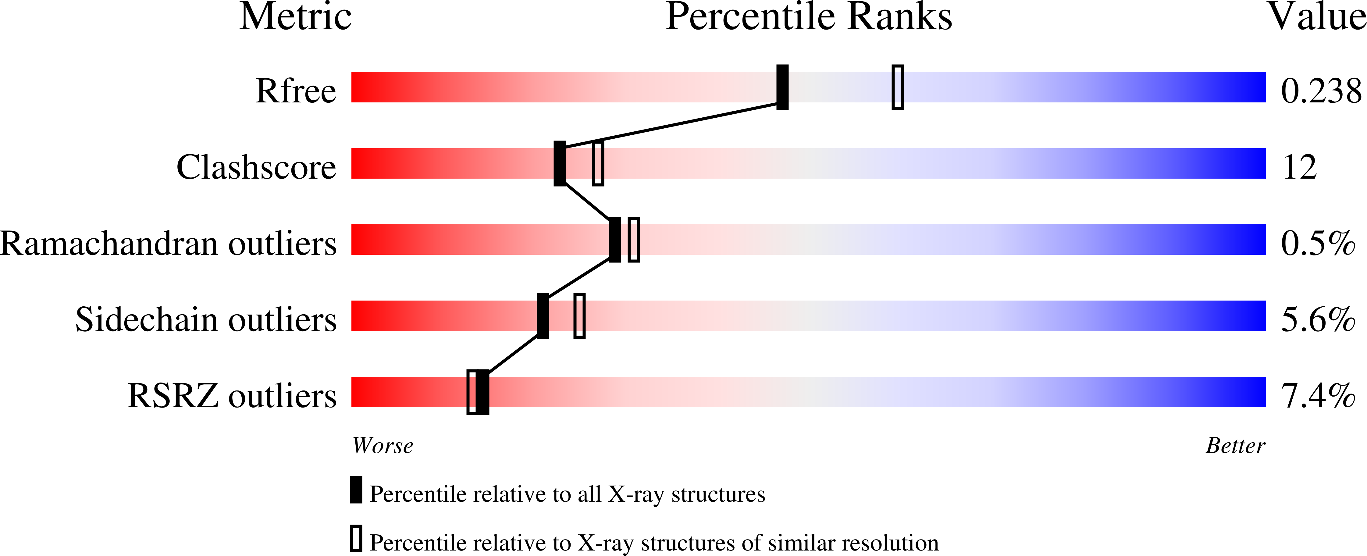
R-Value Free:
0.23
R-Value Work:
0.17
R-Value Observed:
0.18
Space Group:
P 1 21 1