1Y8Z image
Deposition Date 2004-12-14
Release Date 2005-08-30
Last Version Date 2024-11-20
Entry Detail
PDB ID:
1Y8Z
Keywords:
Title:
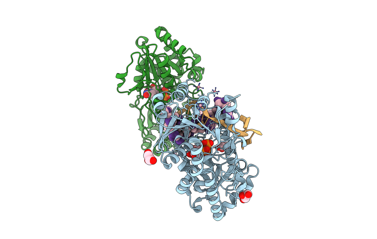
alpha-glucosyltransferase in complex with UDP and a 13-mer DNA containing a HMU base at 1.9 A resolution
Biological Source:
Source Organism:
Host Organism:
Method Details:
Experimental Method:
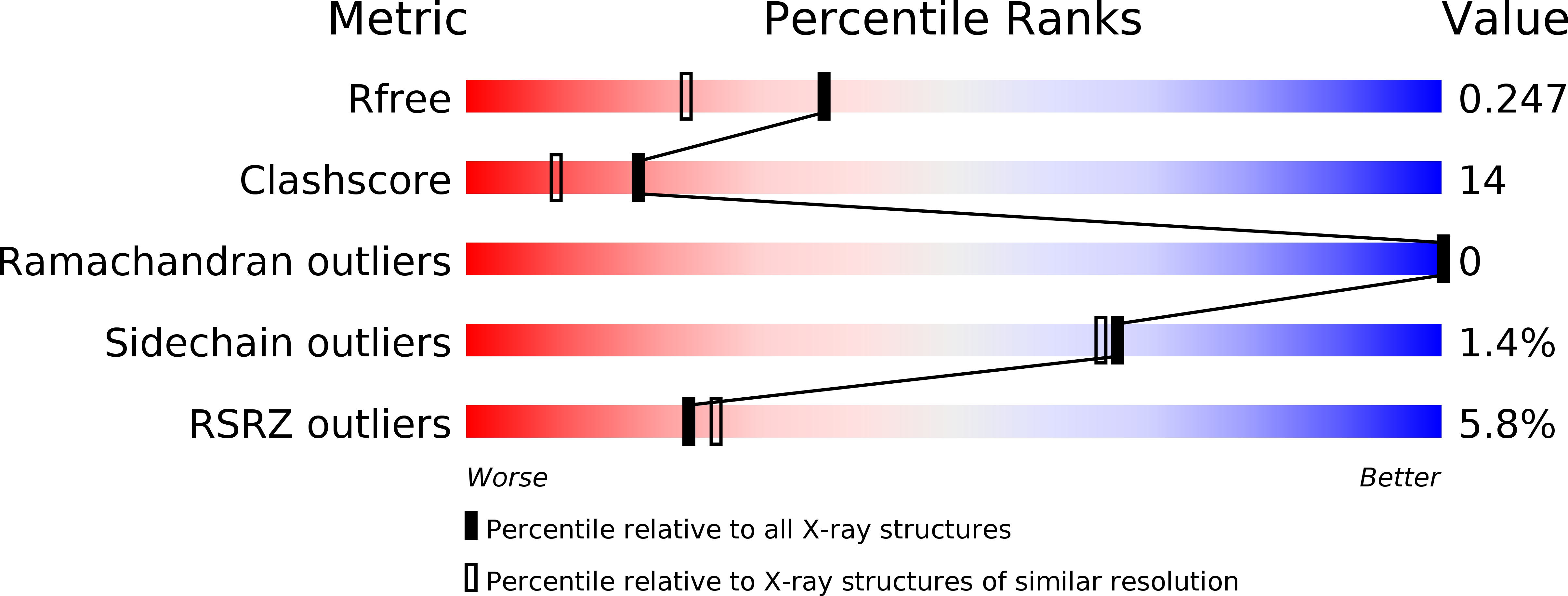
Resolution:
1.90 Å
R-Value Free:
0.25
R-Value Work:
0.20
Space Group:
P 1 21 1
Macromolecular Entities
Structures with similar UniProt ID
Protein Blast
Polymer Type:polypeptide(L)
Molecule:DNA alpha-glucosyltransferase
Gene (Uniprot):agt
Chain IDs:C (auth: A), D (auth: B)
Chain Length:402
Number of Molecules:2
Biological Source:Enterobacteria phage T4
Modified Residue
Compound ID Chain ID Parent Comp ID Details 2D Image
5HU A DU ?
CME C CYS S,S-(2-HYDROXYETHYL)THIOCYSTEINE
Primary Citation
Structural evidence of a passive base-flipping mechanism for AGT, an unusual GT-B glycosyltransferase.
J.Mol.Biol. 352 139 150 (2005)
PMID: 16081100 DOI: 10.1016/j.jmb.2005.07.007

Abstact

The Escherichia coli T4 bacteriophage uses two glycosyltransferases to glucosylate and thus protect its DNA: the retaining alpha-glucosyltransferase (AGT) and the inverting beta-glucosyltransferase (BGT). They glucosylate 5-hydroxymethyl cytosine (5-HMC) bases of duplex DNA using UDP-glucose as the sugar donor to form an alpha-glucosidic linkage and a beta-glucosidic linkage, respectively. Five structures of AGT have been determined: a binary complex with the UDP product and four ternary complexes with UDP or UDP-glucose and oligonucleotides containing an A:G, HMU:G (hydroxymethyl uracyl) or AP:G (apurinic/apyrimidinic) mismatch at the target base-pair. AGT adopts the GT-B fold, one of the two folds known for GTs. However, while the sugar donor binding mode is classical for a GT-B enzyme, the sugar acceptor binding mode is unexpected and breaks the established consensus: AGT is the first GT-B enzyme that predominantly binds both the sugar donor and acceptor to the C-terminal domain. Its active site pocket is highly similar to four retaining GT-B glycosyltransferases (trehalose-6-phosphate synthase, glycogen synthase, glycogen and maltodextrin phosphorylases) strongly suggesting a common evolutionary origin and catalytic mechanism for these enzymes. Structure-guided mutagenesis and kinetic analysis do not permit identification of a nucleophile residue responsible for a glycosyl-enzyme intermediate for the classical double displacement mechanism. Interestingly, the DNA structures reveal partially flipped-out bases. They provide evidence for a passive role of AGT in the base-flipping mechanism and for its specific recognition of the acceptor base.

Legend

Protein

Chemical

Disease

Primary Citation of related structures
Feedback Form
Name
Email
Institute
Feedback