1Y5V image
Entry Detail
PDB ID:
1Y5V
Keywords:
Title:
tRNA-Guanine Transglycosylase (TGT) in complex with 6-Amino-4-(2-phenylethyl)-1,7-dihydro-8H-imidazo[4,5-g]quinazolin-8-one
Biological Source:
Source Organism:
PDB Version:
Deposition Date:
2004-12-03
Release Date:
2005-12-13
Method Details:
Experimental Method:
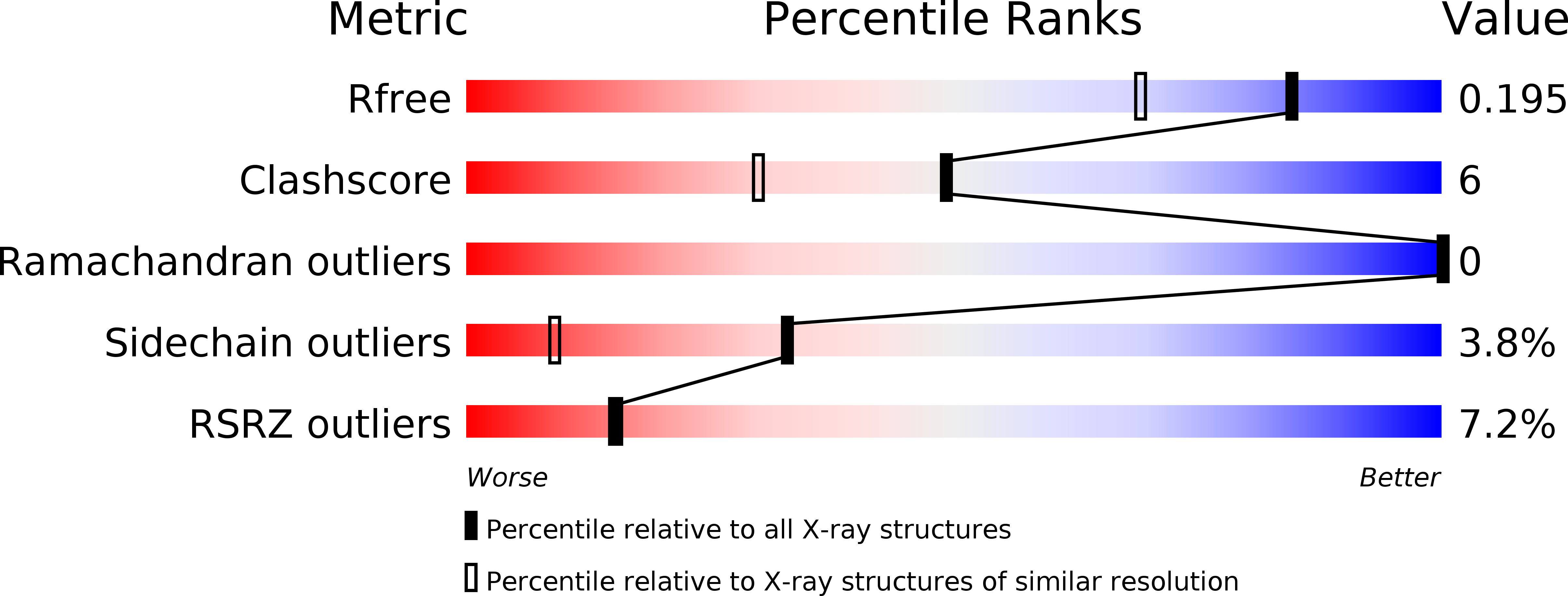
Resolution:
1.58 Å
R-Value Free:
0.21
R-Value Work:
0.16
R-Value Observed:
0.16
Space Group:
C 1 2 1
Macromolecular Entities
Polymer Type:polypeptide(L)
Description:Queuine tRNA-ribosyltransferase
Chain IDs:A
Chain Length:385
Number of Molecules:1
Biological Source:Zymomonas mobilis
Primary Citation
Crystal structures of tRNA-guanine transglycosylase (TGT) in complex with novel and potent inhibitors unravel pronounced induced-fit adaptations and suggest dimer formation upon substrate binding
J.Mol.Biol. 370 492 511 (2007)
PMID: 17524419 DOI: 10.1016/j.jmb.2007.04.008

Abstact

The bacterial tRNA-guanine transglycosylase (TGT) is a tRNA modifying enzyme catalyzing the exchange of guanine 34 by the modified base preQ1. The enzyme is involved in the infection pathway of Shigella, causing bacterial dysentery. As no crystal structure of the Shigella enzyme is available the homologous Zymomonas mobilis TGT was used for structure-based drug design resulting in new, potent, lin-benzoguanine-based inhibitors. Thorough kinetic studies show size-dependent inhibition of these compounds resulting in either a competitive or non-competitive blocking of the base exchange reaction in the low micromolar range. Four crystal structures of TGT-inhibitor complexes were determined with a resolution of 1.58-2.1 A. These structures give insight into the structural flexibility of TGT necessary to perform catalysis. In three of the structures molecular rearrangements are observed that match with conformational changes also noticed upon tRNA substrate binding. Several water molecules are involved in these rearrangement processes. Two of them demonstrate the structural and catalytic importance of water molecules during TGT base exchange reaction. In the fourth crystal structure the inhibitor unexpectedly interferes with protein contact formation and crystal packing. In all presently known TGT crystal structures the enzyme forms tightly associated homodimers internally related by crystallographic symmetry. Upon binding of the fourth inhibitor the dimer interface, however, becomes partially disordered. This result prompted further analyses to investigate the relevance of dimer formation for the functional protein. Consultation of the available TGT structures and sequences from different species revealed structural and functional conservation across the contacting residues. This suggests that bacterial and eukaryotic TGT could possibly act as homodimers in catalysis. It is hypothesized that one unit of the dimer performs the catalytic reaction whereas the second is required to recognize and properly orient the bound tRNA for the catalytic reaction.

Legend

Protein

Chemical

Disease

Primary Citation of related structures