1Y57 image
Deposition Date 2004-12-02
Release Date 2005-06-21
Last Version Date 2024-03-13
Entry Detail
PDB ID:
1Y57
Keywords:
Title:
Structure of unphosphorylated c-Src in complex with an inhibitor
Biological Source:
Source Organism:
Homo sapiens (Taxon ID: 9606)
Host Organism:
Method Details:
Experimental Method:
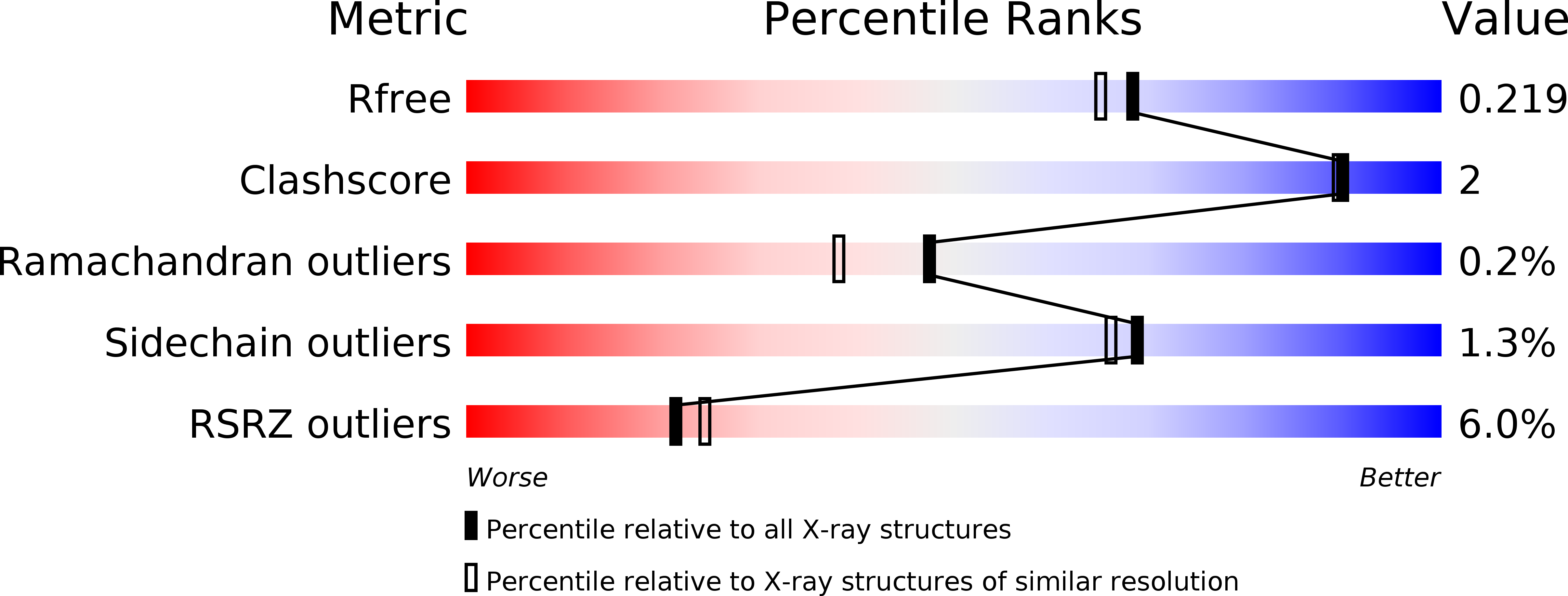
Resolution:
1.91 Å
R-Value Free:
0.21
R-Value Work:
0.18
R-Value Observed:
0.18
Space Group:
P 41 21 2
Macromolecular Entities
Structures with similar UniProt ID
Protein Blast
Polymer Type:polypeptide(L)
Molecule:Proto-oncogene tyrosine-protein kinase Src
Gene (Uniprot):SRC
Chain IDs:A
Chain Length:452
Number of Molecules:1
Biological Source:Homo sapiens
Primary Citation
The Crystal Structure of a c-Src Complex in an Active Conformation Suggests Possible Steps in c-Src Activation
Structure 13 861 871 (2005)
PMID: 15939018 DOI: 10.1016/j.str.2005.03.012

Abstact

The regulation of the activity of Abl and Src family tyrosine kinases is mediated by intramolecular interactions between the SH3, SH2, and kinase (SH1) domains. We have determined the crystal structure of an unphosphorylated form of c-Src in which the SH2 domain is not bound to the C-terminal tail. This results in an open structure where the kinase domain adopts an active conformation and the C terminus binds within a hydrophobic pocket in the C-terminal lobe. NMR binding studies support the hypothesis that an N-terminal myristate could bind in this pocket, as observed for Abl, suggesting that c-Src may also be regulated by myristate binding. In addition, the structure contains a des-methyl analog of the antileukemia drug imatinib (STI571; Gleevec). This structure reveals why the drug shows a low affinity for active kinase conformations, contributing to its excellent kinase selectivity profile.

Legend

Protein

Chemical

Disease

Primary Citation of related structures