1Y4Y image
Deposition Date 2004-12-01
Release Date 2005-02-22
Last Version Date 2024-04-03
Entry Detail
PDB ID:
1Y4Y
Title:
X-ray crystal structure of Bacillus stearothermophilus Histidine phosphocarrier protein (Hpr)
Biological Source:
Source Organism:
Host Organism:
Method Details:
Experimental Method:
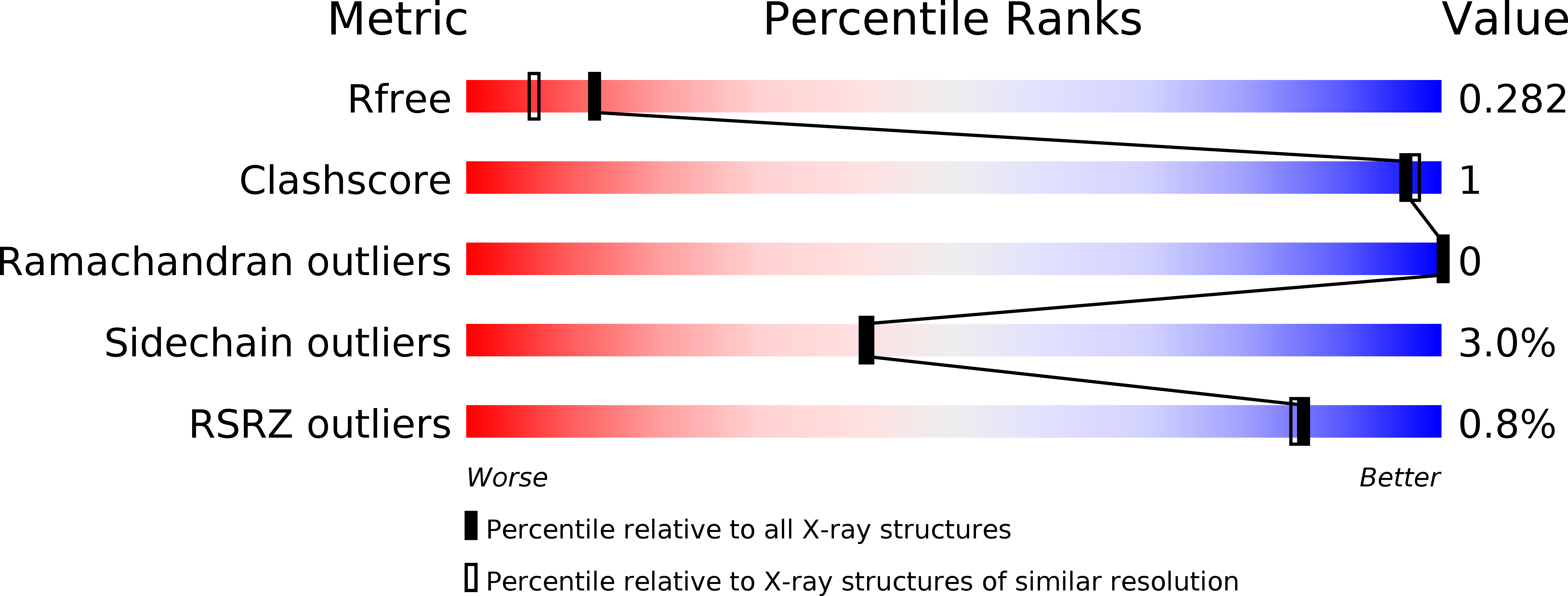
Resolution:
2.00 Å
R-Value Free:
0.28
R-Value Work:
0.21
R-Value Observed:
0.21
Space Group:
I 41 2 2
Macromolecular Entities
Structures with similar UniProt ID
Protein Blast
Polymer Type:polypeptide(L)
Molecule:Phosphocarrier protein HPr
Gene (Uniprot):ptsH
Chain IDs:A, B, C
Chain Length:88
Number of Molecules:3
Biological Source:Geobacillus stearothermophilus
Ligand Molecules
Primary Citation
The HPr proteins from the thermophile Bacillus stearothermophilus can form domain-swapped dimers.
J.Mol.Biol. 346 919 931 (2005)
PMID: 15713472 DOI: 10.1016/j.jmb.2004.12.008

Abstact

The study of proteins from extremophilic organisms continues to generate interest in the field of protein folding because paradigms explaining the enhanced stability of these proteins still elude us and such studies have the potential to further our knowledge of the forces stabilizing proteins. We have undertaken such a study with our model protein HPr from a mesophile, Bacillus subtilis, and a thermophile, Bacillus stearothermophilus. We report here the high-resolution structures of the wild-type HPr protein from the thermophile and a variant, F29W. The variant proved to crystallize in two forms: a monomeric form with a structure very similar to the wild-type protein as well as a domain-swapped dimer. Interestingly, the structure of the domain-swapped dimer for HPr is very different from that observed for a homologous protein, Crh, from B.subtilis. The existence of a domain-swapped dimer has implications for amyloid formation and is consistent with recent results showing that the HPr proteins can form amyloid fibrils. We also characterized the conformational stability of the thermophilic HPr proteins using thermal and solvent denaturation methods and have used the high-resolution structures in an attempt to explain the differences in stability between the different HPr proteins. Finally, we present a detailed analysis of the solution properties of the HPr proteins using a variety of biochemical and biophysical methods.

Legend

Protein

Chemical

Disease

Primary Citation of related structures