Deposition Date
2004-12-01
Release Date
2005-02-08
Last Version Date
2024-11-13
Entry Detail
PDB ID:
1Y4J
Keywords:
Title:
Crystal structure of the paralogue of the human formylglycine generating enzyme
Biological Source:
Source Organism(s):
Homo sapiens (Taxon ID: 9606)
Method Details:
Experimental Method:
Resolution:
1.86 Å
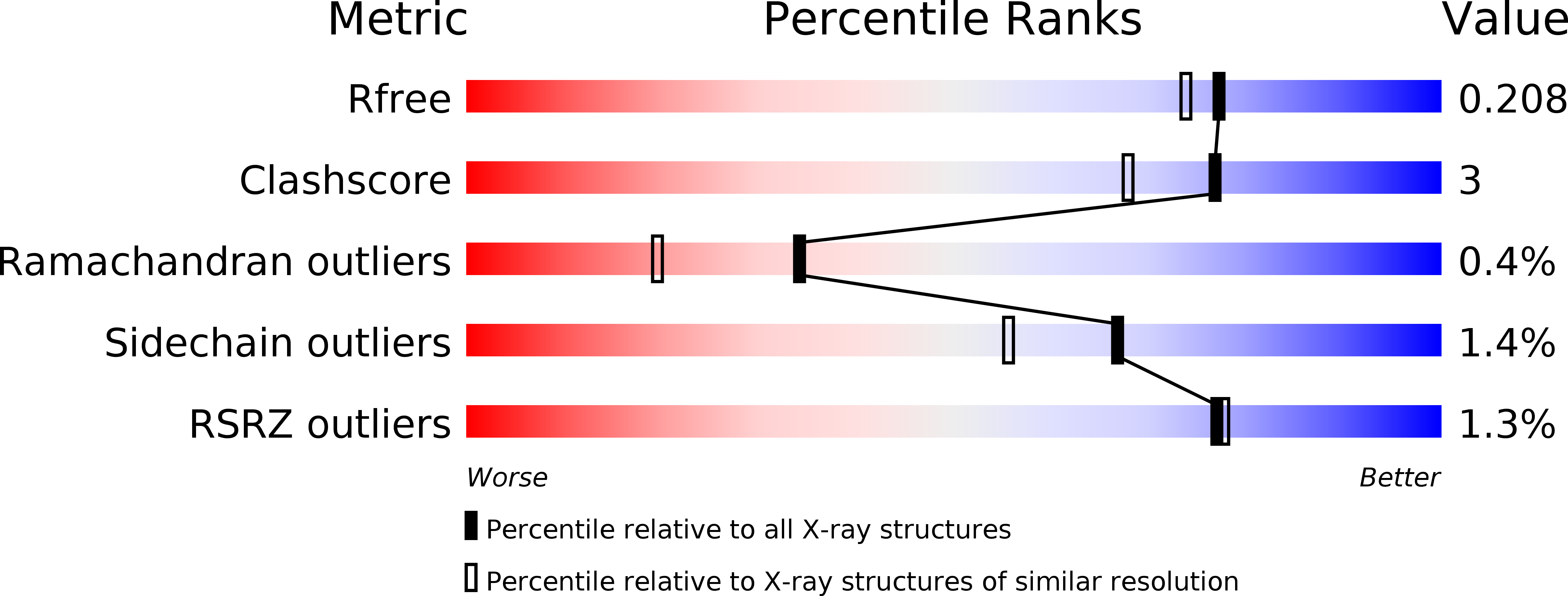
R-Value Free:
0.20
R-Value Work:
0.16
R-Value Observed:
0.17
Space Group:
P 1 21 1