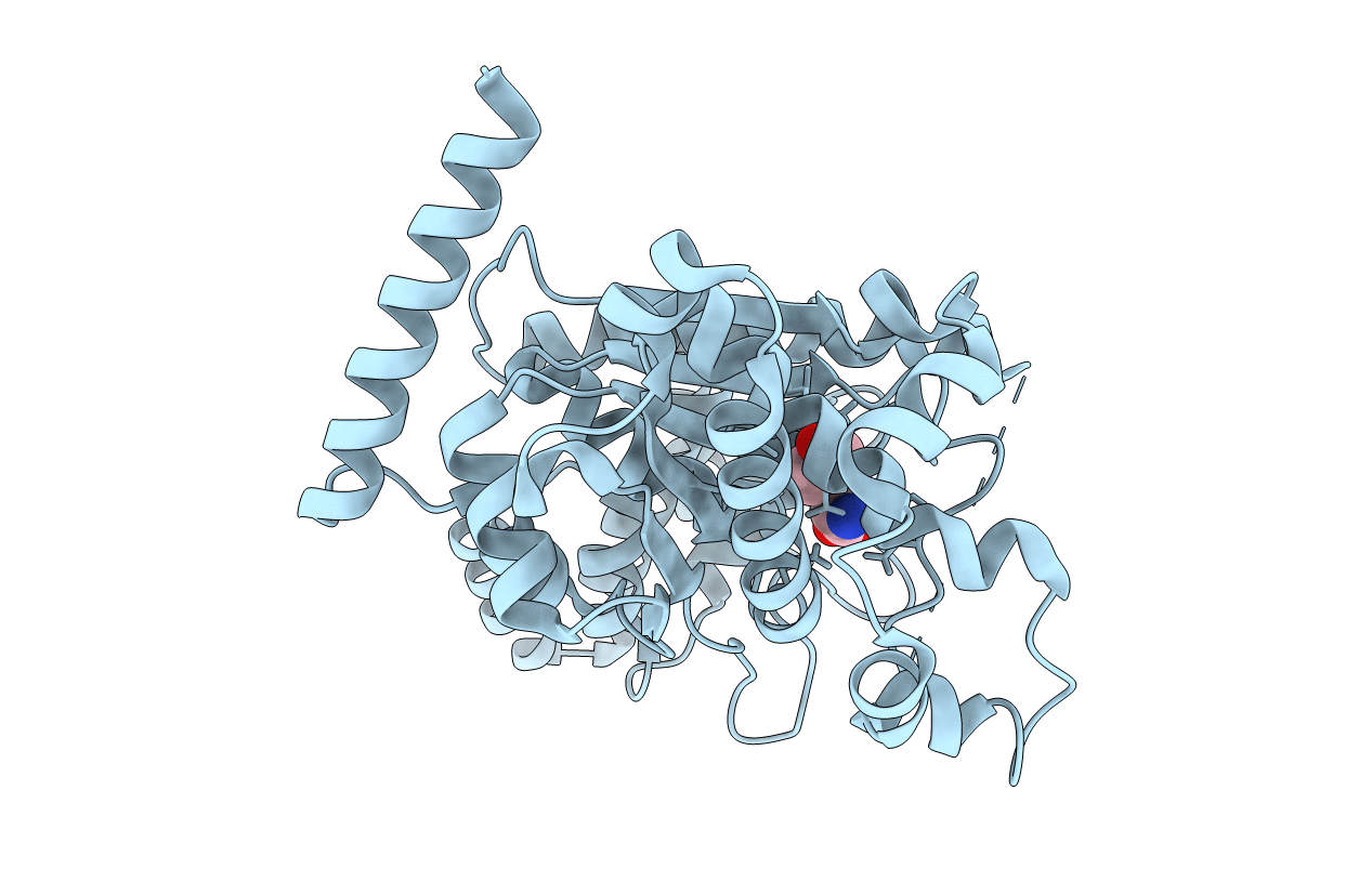
1Y42 image
Entry Detail
PDB ID:
1Y42
Keywords:
Title:
Crystal structure of a C-terminally truncated CYT-18 protein
Biological Source:
Source Organism:
Host Organism:
PDB Version:
Deposition Date:
2004-11-29
Release Date:
2005-02-15
Method Details:
Experimental Method:
Resolution:
1.95 Å
R-Value Free:
0.23
R-Value Work:
0.17
R-Value Observed:
0.17
Space Group:
C 1 2 1
Macromolecular Entities
Polymer Type:polypeptide(L)
Description:Tyrosyl-tRNA synthetase, mitochondrial
Chain IDs:A (auth: X)
Chain Length:392
Number of Molecules:1
Biological Source:Neurospora crassa
Ligand Molecules
Primary Citation
A tyrosyl-tRNA synthetase adapted to function in group I intron splicing by acquiring a new RNA binding surface.
Mol.Cell 17 417 428 (2005)
PMID: 15694342 DOI: 10.1016/j.molcel.2004.12.026

Abstact

We determined a 1.95 A X-ray crystal structure of a C-terminally truncated Neurospora crassa mitochondrial tyrosyl-tRNA synthetase (CYT-18 protein) that functions in splicing group I introns. CYT-18's nucleotide binding fold and intermediate alpha-helical domains superimpose on those of bacterial TyrRSs, except for an N-terminal extension and two small insertions not found in nonsplicing bacterial enzymes. These additions surround the cyt-18-1 mutation site and are sites of suppressor mutations that restore splicing, but not synthetase activity. Highly constrained models based on directed hydroxyl radical cleavage assays show that the group I intron binds at a site formed in part by the three additions on the nucleotide binding fold surface opposite that which binds tRNATyr. Our results show how essential proteins can progressively evolve new functions.

Legend

Protein

Chemical

Disease

Primary Citation of related structures