1Y3T image
Deposition Date 2004-11-26
Release Date 2005-01-18
Last Version Date 2023-08-23
Entry Detail
PDB ID:
1Y3T
Keywords:
Title:
Crystal structure of YxaG, a dioxygenase from Bacillus subtilis
Biological Source:
Source Organism:
Host Organism:
Method Details:
Experimental Method:
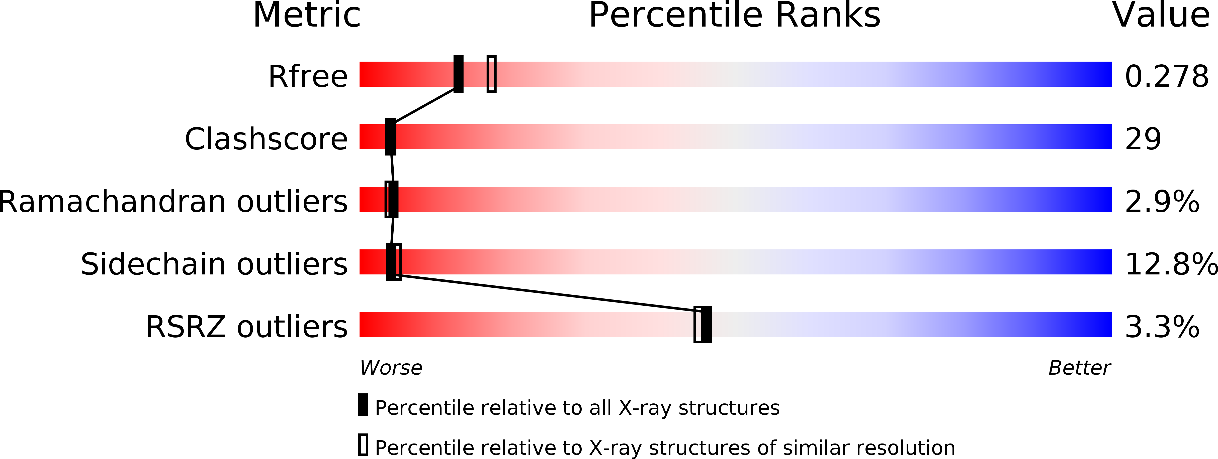
Resolution:
2.40 Å
R-Value Free:
0.27
R-Value Work:
0.21
R-Value Observed:
0.22
Space Group:
P 21 21 2
Macromolecular Entities
Structures with similar UniProt ID
Protein Blast
Polymer Type:polypeptide(L)
Molecule:Hypothetical protein yxaG
Gene (Uniprot):qdoI
Chain IDs:A, B
Chain Length:337
Number of Molecules:2
Biological Source:Bacillus subtilis
Ligand Molecules
Primary Citation
The Crystal Structure of a Quercetin 2,3-Dioxygenase from Bacillus subtilis Suggests Modulation of Enzyme Activity by a Change in the Metal Ion at the Active Site(s)
Biochemistry 44 193 201 (2005)
PMID: 15628860 DOI: 10.1021/bi0484421

Abstact

Common structural motifs, such as the cupin domains, are found in enzymes performing different biochemical functions while retaining a similar active site configuration and structural scaffold. The soil bacterium Bacillus subtilis has 20 cupin genes (0.5% of the total genome) with up to 14% of its genes in the form of doublets, thus making it an attractive system for studying the effects of gene duplication. There are four bicupins in B. subtilis encoded by the genes yvrK, yoaN, yxaG, and ywfC. The gene products of yvrK and yoaN function as oxalate decarboxylases with a manganese ion at the active site(s), whereas YwfC is a bacitracin synthetase. Here we present the crystal structure of YxaG, a novel iron-containing quercetin 2,3-dioxygenase with one active site in each cupin domain. Yxag is a dimer, both in solution and in the crystal. The crystal structure shows that the coordination geometry of the Fe ion is different in the two active sites of YxaG. Replacement of the iron at the active site with other metal ions suggests modulation of enzymatic activity in accordance with the Irving-Williams observation on the stability of metal ion complexes. This observation, along with a comparison with the crystal structure of YvrK determined recently, has allowed for a detailed structure-function analysis of the active site, providing clues to the diversification of function in the bicupin family of proteins.

Legend

Protein

Chemical

Disease

Primary Citation of related structures