1Y3G image
Entry Detail
PDB ID:
1Y3G
Keywords:
Title:
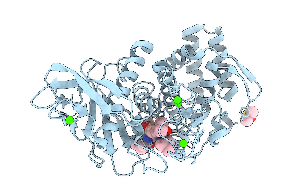
Crystal Structure of a Silanediol Protease Inhibitor Bound to Thermolysin
Biological Source:
PDB Version:
Deposition Date:
2004-11-24
Release Date:
2006-01-17
Method Details:
Experimental Method:
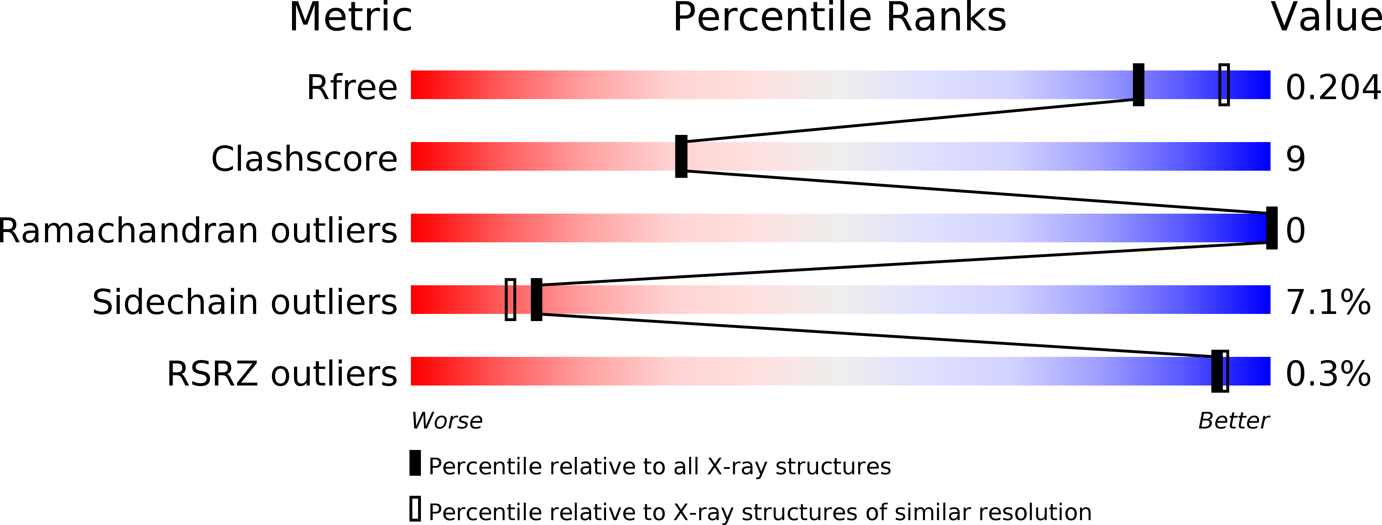
Resolution:
2.10 Å
R-Value Free:
0.22
R-Value Work:
0.15
R-Value Observed:
0.15
Space Group:
P 61 2 2
Macromolecular Entities
Polymer Type:polypeptide(L)
Description:Thermolysin
Chain IDs:A (auth: E)
Chain Length:316
Number of Molecules:1
Biological Source:Bacillus thermoproteolyticus
Primary Citation
Structural Analysis of Silanediols as Transition-State-Analogue Inhibitors of the Benchmark Metalloprotease Thermolysin(,).
Biochemistry 44 16524 16528 (2005)
PMID: 16342943 DOI: 10.1021/bi051346v

Abstact

Dialkylsilanediols have been found to be an effective functional group for the design of active-site-directed protease inhibitors, including aspartic (HIV protease) and metallo (ACE and thermolysin) proteases. The use of silanediols is predicated on its resemblance to the hydrated carbonyl transition-state structure of amide hydrolysis. This concept has been tested by replacing the presumed tetrahedral carbon of a thermolysin substrate with a silanediol group, resulting in an inhibitor with an inhibition constant K(i) = 40 nM. The structure of the silanediol bound to the active site of thermolysin was found to have a conformation very similar to that of a corresponding phosphonamidate inhibitor (K(i) = 10 nM). In both cases, a single oxygen is within bonding distance to the active-site zinc ion, mimicking the presumed tetrahedral transition state. There are binding differences that appear to be related to the presence or absence of protons on the oxygens attached to the silicon or phosphorus. This is the first crystal structure of an organosilane bound to the active site of a protease.

Legend

Protein

Chemical

Disease

Primary Citation of related structures