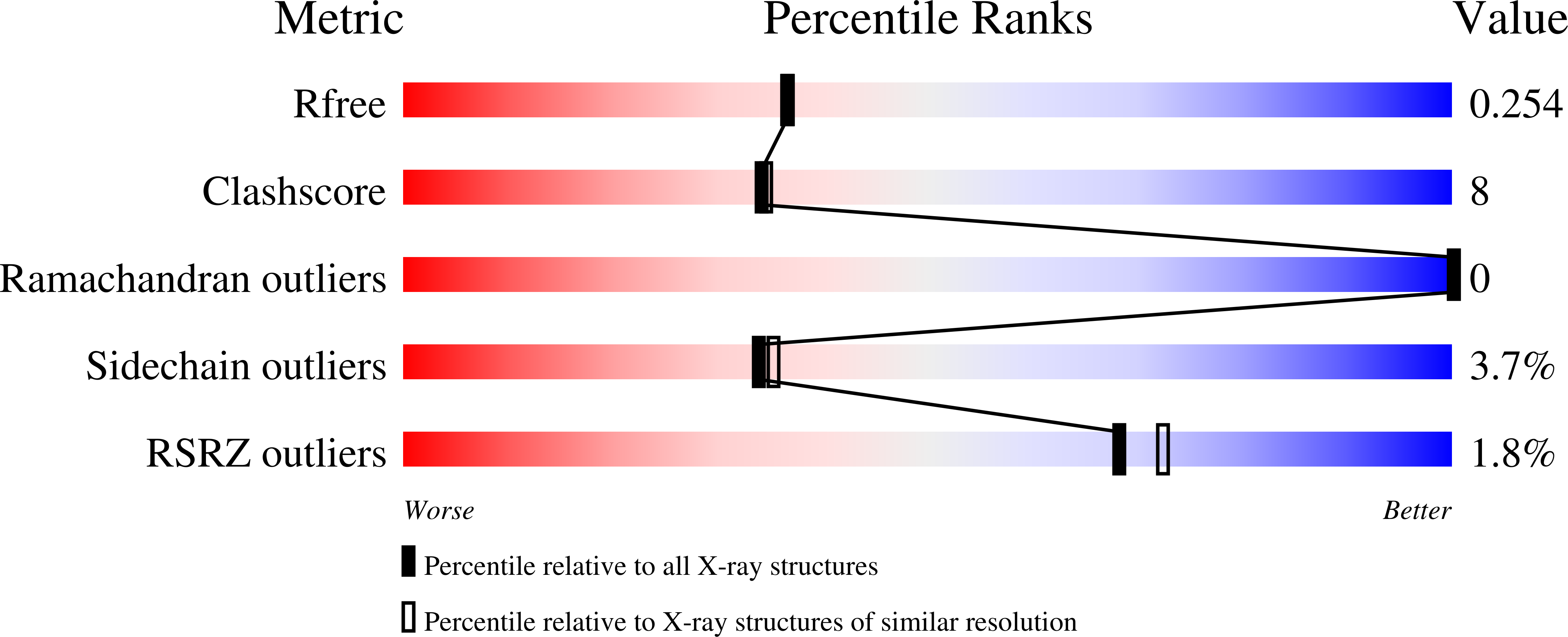
1Y25 image
Deposition Date 2004-11-20
Release Date 2005-11-20
Last Version Date 2023-10-25
Entry Detail
PDB ID:
1Y25
Keywords:
Title:
structure of mycobacterial thiol peroxidase Tpx
Biological Source:
Source Organism:
Host Organism:
Method Details:
Experimental Method:
Resolution:
2.10 Å
R-Value Free:
0.24
R-Value Work:
0.17
R-Value Observed:
0.17
Space Group:
P 31 2 1
Macromolecular Entities
Polymer Type:polypeptide(L)
Molecule:Probable thiol peroxidase
Mutations:C60S
Chain IDs:A, B
Chain Length:165
Number of Molecules:2
Biological Source:Mycobacterium tuberculosis
Ligand Molecules
Primary Citation
Structure of the inactive variant C60S of Mycobacterium tuberculosis thiol peroxidase
Acta Crystallogr.,Sect.D 62 563 567 (2006)
PMID: 16627951 DOI: 10.1107/S0907444906008249

Abstact

The genome of Mycobacterium tuberculosis encodes several peroxiredoxins (Prxs) thought to be active against organic and inorganic peroxides. The open reading frame Rv1932 encodes a 165-residue thiol peroxidase (Tpx), which belongs to the atypical 2-Cys peroxiredoxin family. The crystal structure of the C60S mutant of M. tuberculosis Tpx (MtTpx) crystallized in space group P3(1)21, with unit-cell parameters a = 106.08, b = 106.08, c = 65.33 A. The structure has been refined to an R value of 17.1% (R(free) = 24.9%) at 2.1 A resolution. MtTpx is structurally homologous to other peroxiredoxins, including the mycobacterial AhpC and AhpE. The inactive MtTpx C60S mutant structure closely resembles the structure of Streptococcus pneumoniae Tpx (SpTpx) and thus represents the reduced enzyme state. The mutated active-site serine is electrostatically linked to Arg130 and hydrogen bonded to Thr57, practically identical to the cysteine in SpTpx. A cocrystallized acetate molecule mimics the position of the substrate and interacts with Ser60, Arg130 and Thr57.

Legend

Protein

Chemical

Disease

Primary Citation of related structures