1Y0Q image
Deposition Date 2004-11-16
Release Date 2004-12-21
Last Version Date 2024-02-14
Entry Detail
PDB ID:
1Y0Q
Keywords:
Title:
Crystal structure of an active group I ribozyme-product complex
Biological Source:
Source Organism:
Host Organism:
Method Details:
Experimental Method:
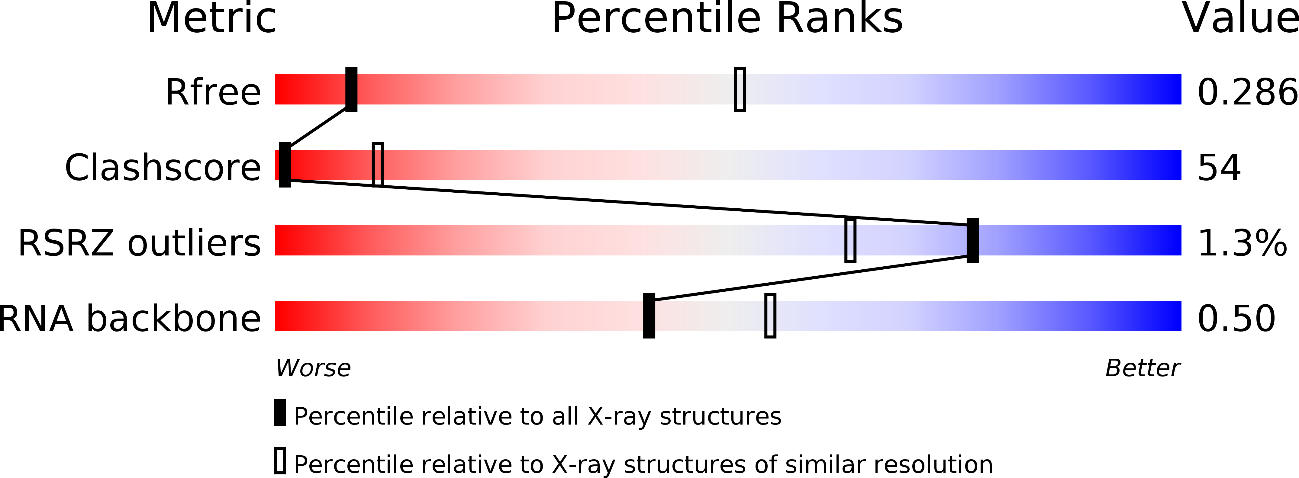
Resolution:
3.60 Å
R-Value Free:
0.31
R-Value Work:
0.27
R-Value Observed:
0.27
Space Group:
I 21 21 21
Macromolecular Entities
Polymer Type:polyribonucleotide
Molecule:Group I ribozyme
Chain IDs:A
Chain Length:229
Number of Molecules:1
Biological Source:Staphylococcus phage Twort
Polymer Type:polyribonucleotide
Molecule:5'-R(*GP*CP*UP*U)-3'
Chain IDs:B
Chain Length:4
Number of Molecules:1
Biological Source:
Primary Citation
Crystal structure of a phage Twort group I ribozyme-product complex
Nat.Struct.Mol.Biol. 12 82 89 (2005)
PMID: 15580277 DOI: 10.1038/nsmb868

Abstact

Group I introns are catalytic RNAs capable of orchestrating two sequential phosphotransesterification reactions that result in self-splicing. To understand how the group I intron active site facilitates catalysis, we have solved the structure of an active ribozyme derived from the orf142-I2 intron from phage Twort bound to a four-nucleotide product RNA at a resolution of 3.6 A. In addition to the three conserved domains characteristic of all group I introns, the Twort ribozyme has peripheral insertions characteristic of phage introns. These elements form a ring that completely envelops the active site, where a snug pocket for guanosine is formed by a series of stacked base triples. The structure of the active site reveals three potential binding sites for catalytic metals, and invokes a role for the 2' hydroxyl of the guanosine substrate in organization of the active site for catalysis.

Legend

Protein

Chemical

Disease

Primary Citation of related structures