Deposition Date
2004-11-13
Release Date
2005-01-25
Last Version Date
2024-02-14
Entry Detail
PDB ID:
1XZZ
Keywords:
Title:
Crystal structure of the ligand binding suppressor domain of type 1 inositol 1,4,5-trisphosphate receptor
Biological Source:
Source Organism:
Mus musculus (Taxon ID: 10090)
Host Organism:
Method Details:
Experimental Method:
Resolution:
1.80 Å
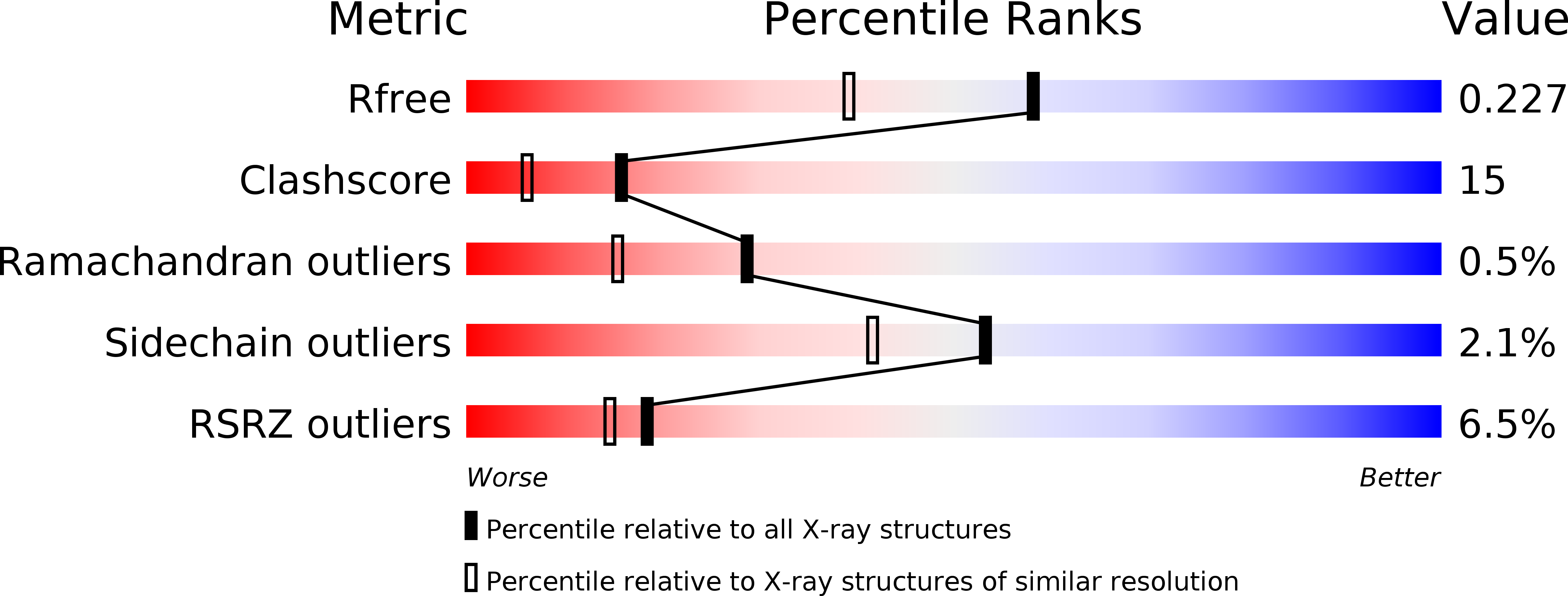
R-Value Free:
0.23
R-Value Work:
0.20
R-Value Observed:
0.20
Space Group:
H 3 2