1XYI image
Deposition Date 2004-11-10
Release Date 2005-02-08
Last Version Date 2024-05-29
Entry Detail
PDB ID:
1XYI
Title:
Hyperthermophile chromosomal protein Sac7d double mutant Val26Ala/Met29Ala in complex with DNA GCGATCGC
Biological Source:
Source Organism:
Host Organism:
Method Details:
Experimental Method:
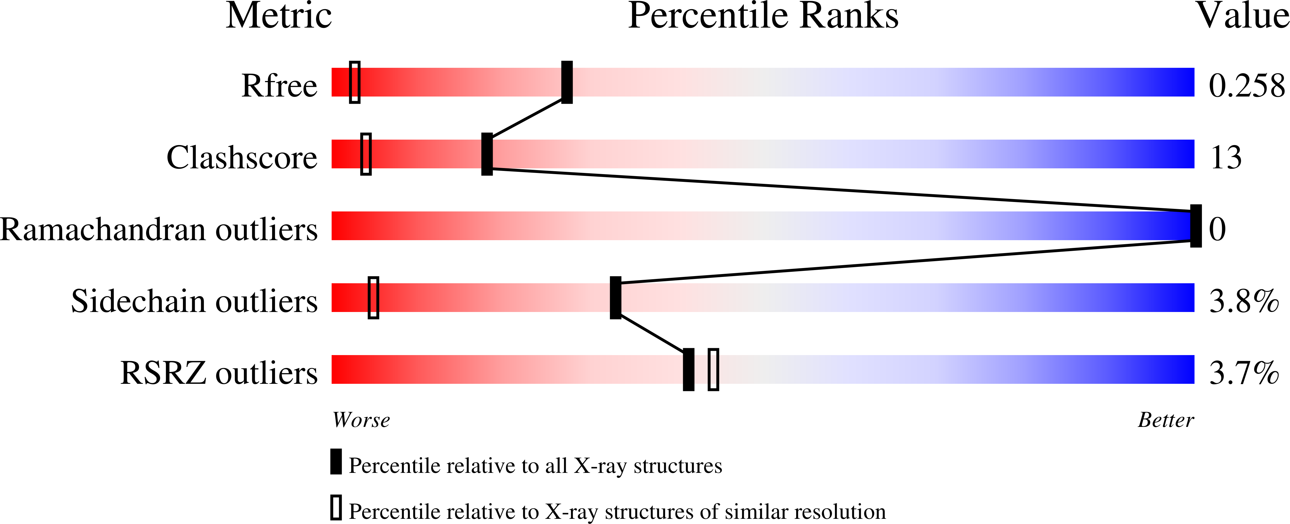
Resolution:
1.45 Å
R-Value Free:
0.25
R-Value Work:
0.21
R-Value Observed:
0.21
Space Group:
P 21 21 21
Macromolecular Entities
Structures with similar UniProt ID
Protein Blast
Polymer Type:polypeptide(L)
Molecule:DNA-binding proteins 7a/7b/7d
Gene (Uniprot):Saci_0064
Mutagens:V26A, M29A
Chain IDs:C (auth: A)
Chain Length:66
Number of Molecules:1
Biological Source:Sulfolobus acidocaldarius
Primary Citation
Probing the DNA kink structure induced by the hyperthermophilic chromosomal protein Sac7d
Nucleic Acids Res. 33 430 438 (2005)
PMID: 15653643 DOI: 10.1093/nar/gki191

Abstact

Sac7d, a small, abundant, sequence-general DNA-binding protein from the hyperthermophilic archaeon Sulfolobus acidocaldarius, causes a single-step sharp kink in DNA (approximately 60 degrees) via the intercalation of both Val26 and Met29. These two amino acids were systematically changed in size to probe their effects on DNA kinking. Eight crystal structures of five Sac7d mutant-DNA complexes have been analyzed. The DNA-binding pattern of the V26A and M29A single mutants is similar to that of the wild-type, whereas the V26A/M29A protein binds DNA without side chain intercalation, resulting in a smaller overall bending (approximately 50 degrees). The M29F mutant inserts the Phe29 side chain orthogonally to the C2pG3 step without stacking with base pairs, inducing a sharp kink (approximately 80 degrees). In the V26F/M29F-GCGATCGC complex, Phe26 intercalates deeply into DNA bases by stacking with the G3 base, whereas Phe29 is stacked on the G15 deoxyribose, in a way similar to those used by the TATA box-binding proteins. All mutants have reduced DNA-stabilizing ability, as indicated by their lower T m values. The DNA kink patterns caused by different combinations of hydrophobic side chains may be relevant in understanding the manner by which other minor groove-binding proteins interact with DNA.

Legend

Protein

Chemical

Disease

Primary Citation of related structures
Feedback Form
Name
Email
Institute
Feedback