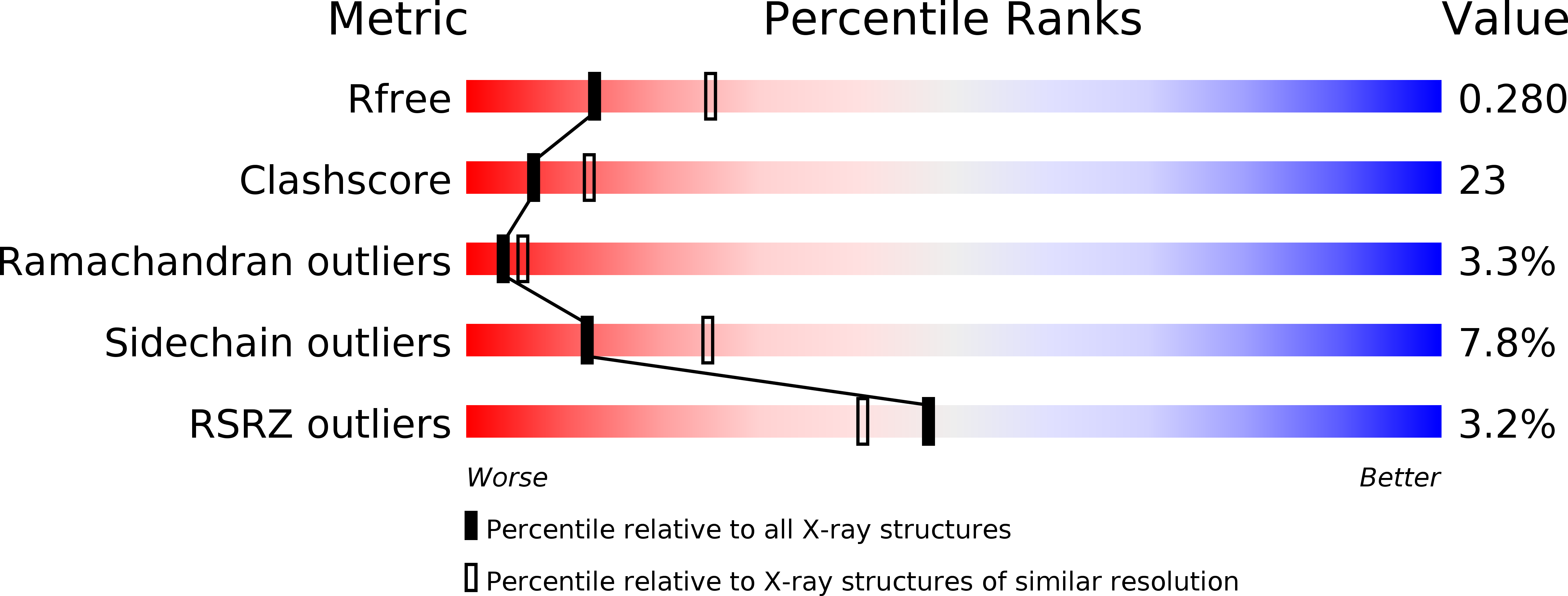
1XWJ image
Deposition Date 2004-11-01
Release Date 2005-10-11
Last Version Date 2024-03-13
Entry Detail
PDB ID:
1XWJ
Title:
Vinculin head (1-258) in complex with the talin vinculin binding site 3 (1945-1969)
Biological Source:
Source Organism:
Gallus gallus (Taxon ID: 9031)
(Taxon ID: )
Method Details:
Experimental Method:
Resolution:
2.60 Å
R-Value Free:
0.28
R-Value Work:
0.24
Space Group:
P 21 21 2
Macromolecular Entities
Polymer Type:polypeptide(L)
Molecule:Vinculin
Gene (Uniprot):VCL
Chain IDs:A
Chain Length:280
Number of Molecules:1
Biological Source:Gallus gallus
Polymer Type:polypeptide(L)
Molecule:Talin
Chain IDs:B
Chain Length:26
Number of Molecules:1
Biological Source:
Primary Citation
Structural and Dynamic Characterization of a Vinculin Binding Site in the Talin Rod
Biochemistry 45 1805 1817 (2006)
PMID: 16460027 DOI: 10.1021/bi052136l

Abstact

Talin is a key protein involved in linking integrins to the actin cytoskeleton. The long flexible talin rod domain contains a number of binding sites for vinculin, a cytoskeletal protein important in stabilizing integrin-mediated cell-matrix junctions. Here we report the solution structure of a talin rod polypeptide (residues 1843-1973) which contains a single vinculin binding site (VBS; residues 1944-1969). Like other talin rod polypeptides, it consists of a helical bundle, in this case a four-helix bundle with a right-handed topology. The residues in the VBS important for vinculin binding were identified by studying the binding of a series of VBS-related peptides to the vinculin Vd1 domain. The key binding determinants are buried in the interior of the helical bundle, suggesting that a substantial structural change in the talin polypeptide is required for vinculin binding. Direct evidence for this was obtained by NMR and EPR spectroscopy. [1H,15N]-HSQC spectra of the talin fragment indicate that vinculin binding caused approximately two-thirds of the protein to adopt a flexible random coil. For EPR spectroscopy, nitroxide spin labels were attached to the talin polypeptide via appropriately located cysteine residues. Measurements of inter-nitroxide distances in doubly spin-labeled protein showed clearly that the helical bundle is disrupted and the mobility of the helices, except for the VBS helix, is markedly increased. Binding of vinculin to talin is thus a clear example of the unusual phenomenon of protein unfolding being required for protein/protein interaction.

Legend

Protein

Chemical

Disease

Primary Citation of related structures