Planned Maintenance: Some services may turn out to be unavailable from 15th January, 2026 to 16th January, 2026. We apologize for the inconvenience!

1XWD image
Deposition Date 2004-10-30
Release Date 2005-01-25
Last Version Date 2024-12-25
Entry Detail
PDB ID:
1XWD
Title:
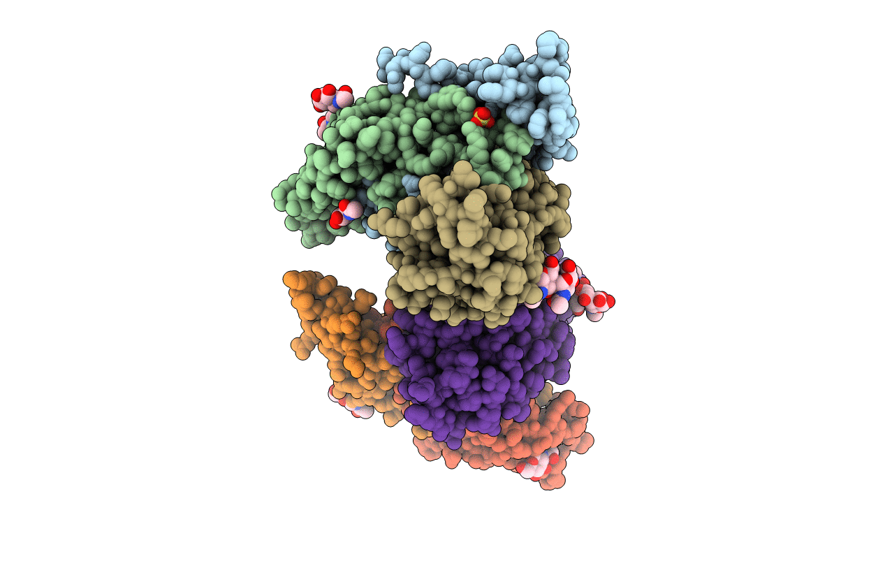
Crystal Structure of Human Follicle Stimulating Hormone Complexed with its Receptor
Biological Source:
Source Organism(s):
Homo sapiens (Taxon ID: 9606)
Expression System(s):
Method Details:
Experimental Method:
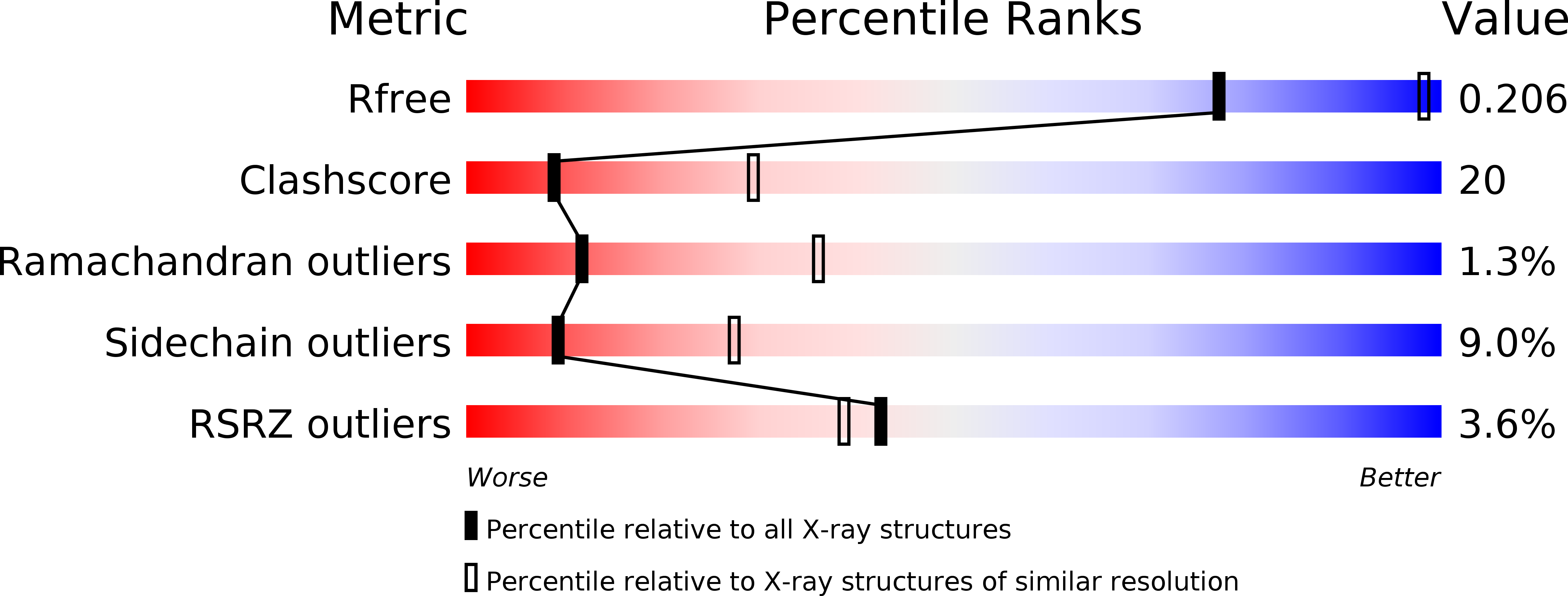
Resolution:
2.92 Å
R-Value Free:
0.25
R-Value Work:
0.21
R-Value Observed:
0.22
Space Group:
C 1 2 1
Macromolecular Entities
Structures with similar UniProt ID
Protein Blast
Polymer Type:polypeptide(L)
Molecule:Glycoprotein hormones alpha chain
Gene (Uniprot):CGA
Chain IDs:A, D
Chain Length:92
Number of Molecules:2
Biological Source:Homo sapiens
Structures with similar UniProt ID
Protein Blast
Polymer Type:polypeptide(L)
Molecule:Follitropin beta chain
Gene (Uniprot):FSHB
Chain IDs:B, E
Chain Length:111
Number of Molecules:2
Biological Source:Homo sapiens
Structures with similar UniProt ID
Protein Blast
Polymer Type:polypeptide(L)
Molecule:Follicle stimulating hormone receptor
Gene (Uniprot):FSHR
Chain IDs:C, F
Chain Length:252
Number of Molecules:2
Biological Source:Homo sapiens
Modified Residue
Compound ID Chain ID Parent Comp ID Details 2D Image
ASN A ASN GLYCOSYLATION SITE
Primary Citation
Structure of human follicle-stimulating hormone in complex with its receptor.
Nature 433 269 277 (2005)
PMID: 15662415 DOI: 10.1038/nature03206

Abstact

Follicle-stimulating hormone (FSH) is central to reproduction in mammals. It acts through a G-protein-coupled receptor on the surface of target cells to stimulate testicular and ovarian functions. We present here the 2.9-A-resolution structure of a partially deglycosylated complex of human FSH bound to the extracellular hormone-binding domain of its receptor (FSHR(HB)). The hormone is bound in a hand-clasp fashion to an elongated, curved receptor. The buried interface of the complex is large (2,600 A2) and has a high charge density. Our analysis suggests that all glycoprotein hormones bind to their receptors in this mode and that binding specificity is mediated by key interaction sites involving both the common alpha- and hormone-specific beta-subunits. On binding, FSH undergoes a concerted conformational change that affects protruding loops implicated in receptor activation. The FSH-FSHR(HB) complexes form dimers in the crystal and at high concentrations in solution. Such dimers may participate in transmembrane signal transduction.

Legend

Protein

Chemical

Disease

Primary Citation of related structures
Feedback Form
Name
Email
Institute
Feedback