1XVY image
Deposition Date 2004-10-28
Release Date 2004-12-14
Last Version Date 2023-08-23
Entry Detail
PDB ID:
1XVY
Title:
Crystal Structure of iron-free Serratia marcescens SfuA
Biological Source:
Source Organism:
Host Organism:
Method Details:
Experimental Method:
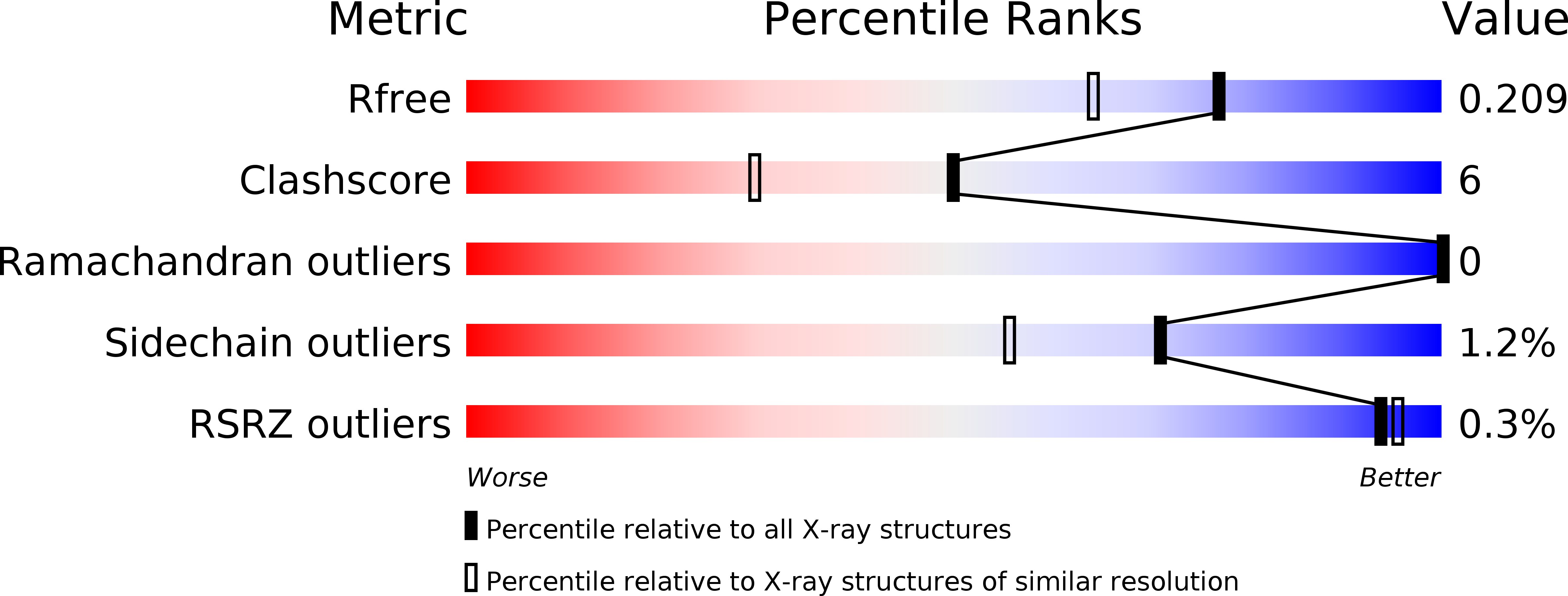
Resolution:
1.74 Å
R-Value Free:
0.20
R-Value Work:
0.16
R-Value Observed:
0.16
Space Group:
P 1 21 1
Macromolecular Entities
Polymer Type:polypeptide(L)
Molecule:sfuA
Gene (Uniprot):fbpA
Chain IDs:A
Chain Length:309
Number of Molecules:1
Biological Source:Yersinia enterocolitica
Ligand Molecules
Primary Citation
Novel Anion-independent Iron Coordination by Members of a Third Class of Bacterial Periplasmic Ferric Ion-binding Proteins
J.Biol.Chem. 280 5820 5827 (2005)
PMID: 15576371 DOI: 10.1074/jbc.M411238200

Abstact

The uptake of the element iron is vital for the survival of most organisms. Numerous pathogenic Gram-negative bacteria utilize a periplasm-to-cytosol ATP-binding cassette transport pathway to transport this essential atom in to the cell. In this study, we investigated the Yersinia enterocolitica (YfuA) and Serratia marcescens (SfuA) iron-binding periplasmic proteins. We have determined the 1.8-angstroms structures of iron-loaded (YfuA) and iron-free (SfuA) forms of this class of proteins. Although the sequence of these proteins varies considerably from the other members of the transferrin structural superfamily, they adopt the same three-dimensional fold. The iron-loaded YfuA structure illustrates the unique nature of this new class of proteins in that they are able to octahedrally coordinate the ferric ion in the absence of a bound anion. The iron-free SfuA structure contains a bound citrate anion in the iron-binding cleft that tethers the N- and C-terminal domains of the apo protein and stabilizes the partially open structure.

Legend

Protein

Chemical

Disease

Primary Citation of related structures