1XVT image
Deposition Date 2004-10-28
Release Date 2005-03-15
Last Version Date 2024-10-30
Entry Detail
PDB ID:
1XVT
Keywords:
Title:
Crystal Structure of Native CaiB in complex with coenzyme A
Biological Source:
Source Organism:
Escherichia coli (Taxon ID: 562)
Host Organism:
Method Details:
Experimental Method:
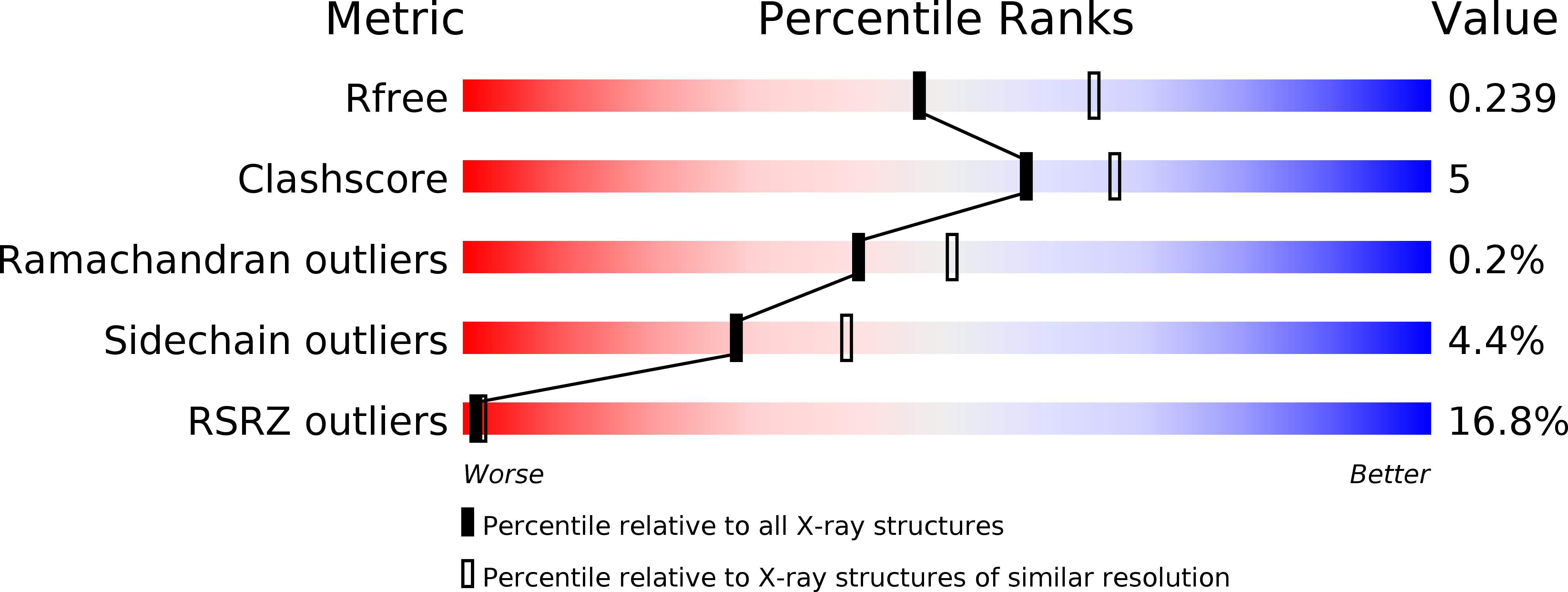
Resolution:
2.30 Å
R-Value Free:
0.23
R-Value Work:
0.19
R-Value Observed:
0.19
Space Group:
P 41 21 2
Macromolecular Entities
Polymer Type:polypeptide(L)
Molecule:Crotonobetainyl-CoA:carnitine CoA-transferase
Gene (Uniprot):caiB
Chain IDs:A
Chain Length:408
Number of Molecules:1
Biological Source:Escherichia coli
Modified Residue
Compound ID Chain ID Parent Comp ID Details 2D Image
MSE A MET SELENOMETHIONINE
Ligand Molecules
Primary Citation
Crystal Structure of Escherichia coli Crotonobetainyl-CoA: Carnitine CoA-Transferase (CaiB) and Its Complexes with CoA and Carnitinyl-CoA.
Biochemistry 44 5728 5738 (2005)
PMID: 15823031 DOI: 10.1021/bi047656f

Abstact

L-Carnitine (R-[-]-3-hydroxy-4-trimethylaminobutyrate) is found in both eukaryotic and prokaryotic cells and participates in diverse processes including long-chain fatty-acid transport and osmoprotection. The enzyme crotonobetainyl/gamma-butyrobetainyl-CoA:carnitine CoA-transferase (CaiB; E.C. 2.8.3.-) catalyzes the first step in carnitine metabolism, leading to the final product gamma-butyrobetaine. The crystal structures of Escherichia coli apo-CaiB, as well as its Asp169Ala mutant bound to CoA and to carnitinyl-CoA, have been determined and refined to 1.6, 2.4, and 2.4 A resolution, respectively. CaiB is composed of two identical circular chains that together form an intertwined dimer. Each monomer consists of a large domain, containing a Rossmann fold, and a small domain. The monomer and dimer resemble those of formyl-CoA transferase from Oxalobacter formigenes, as well as E. coli YfdW, a putative type-III CoA transferase of unknown function. The CoA cofactor-binding site is formed at the interface of the large domain of one monomer and the small domain from the second monomer. Most of the protein-CoA interactions are formed with the Rossmann fold domain. While the location of cofactor binding is similar in the three proteins, the specific CoA-protein interactions vary somewhat between CaiB, formyl-CoA transferase, and YfdW. CoA binding results in a change in the relative positions of the large and small domains compared with apo-CaiB. The observed carnitinyl-CoA product in crystals of the CaiB Asp169Ala mutant cocrystallized with crotonoyl-CoA and carnitine could result from (i) a catalytic mechanism involving a ternary enzyme-substrate complex, independent of a covalent anhydride intermediate with Asp169, (ii) a spontaneous reaction of the substrates in solution, followed by binding to the enzyme, or (iii) an involvement of another residue substituting functionally for Asp169, such as Glu23.

Legend

Protein

Chemical

Disease

Primary Citation of related structures