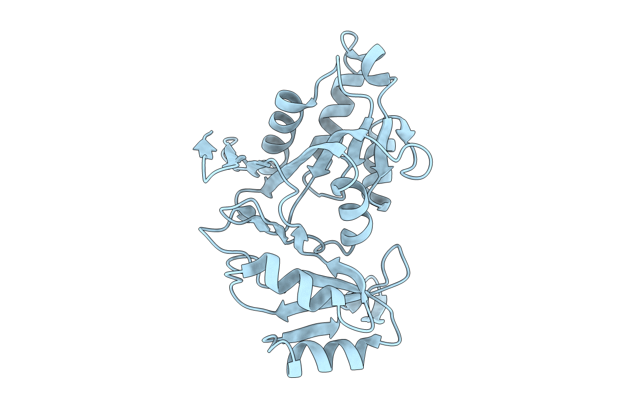
1XTZ image
Deposition Date 2004-10-25
Release Date 2005-08-30
Last Version Date 2023-08-23
Entry Detail
PDB ID:
1XTZ
Keywords:
Title:
Crystal structure of the S. cerevisiae D-ribose-5-phosphate isomerase: comparison with the archeal and bacterial enzymes
Biological Source:
Source Organism:
Host Organism:
Method Details:
Experimental Method:
Resolution:
2.10 Å
R-Value Free:
0.24
R-Value Work:
0.19
R-Value Observed:
0.19
Space Group:
F 4 3 2
Macromolecular Entities
Structures with similar UniProt ID
Protein Blast
Polymer Type:polypeptide(L)
Molecule:Ribose-5-phosphate isomerase
Gene (Uniprot):RKI1
Chain IDs:A
Chain Length:264
Number of Molecules:1
Biological Source:Saccharomyces cerevisiae
Primary Citation
Crystal structure of the S. cerevisiae D-ribose-5-phosphate isomerase: comparison with the archaeal and bacterial enzymes
Biochimie 87 763 769 (2005)
PMID: 16054529 DOI: 10.1016/j.biochi.2005.03.001

Abstact

Ribose-5-phosphate isomerase A has an important role in sugar metabolism by interconverting ribose-5-phosphate and ribulose-5-phosphate. This enzyme is ubiquitous and highly conserved among the three kingdoms of life. We have solved the 2.1 A resolution crystal structure of the Saccharomyces cerevisiae enzyme by molecular replacement. This protein adopts the same fold as its archaeal and bacterial orthologs with two alpha/beta domains tightly packed together. Mapping of conserved residues at the surface of the protein reveals strong invariability of the active site pocket, suggesting a common ligand binding mode and a similar catalytic mechanism. The yeast enzyme associates as a homotetramer similarly to the archaeal protein. The effect of an inactivating mutation (Arg189 to Lys) is discussed in view of the information brought by this structure.

Legend

Protein

Chemical

Disease

Primary Citation of related structures