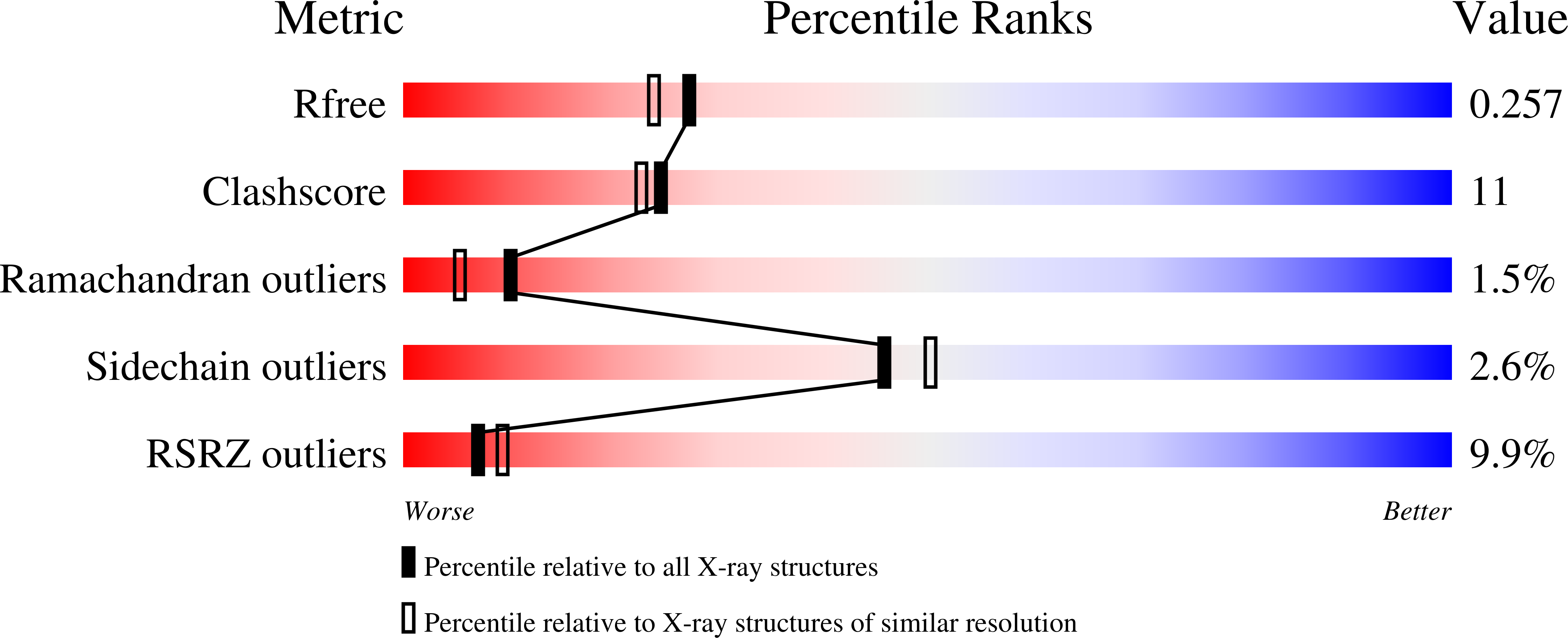
1XTG image
Deposition Date 2004-10-21
Release Date 2004-12-21
Last Version Date 2024-02-14
Entry Detail
PDB ID:
1XTG
Keywords:
Title:
Crystal structure of NEUROTOXIN BONT/A complexed with Synaptosomal-associated protein 25
Biological Source:
Source Organism:
Host Organism:
Method Details:
Experimental Method:
Resolution:
2.10 Å
R-Value Free:
0.24
R-Value Work:
0.21
R-Value Observed:
0.21
Space Group:
P 43 21 2
Macromolecular Entities
Structures with similar UniProt ID
Protein Blast
Polymer Type:polypeptide(L)
Molecule:NEUROTOXIN BONT/A
Mutagens:E224Q, Y366F
Chain IDs:A
Chain Length:424
Number of Molecules:1
Biological Source:Clostridium botulinum
Structures with similar UniProt ID
Protein Blast
Polymer Type:polypeptide(L)
Molecule:Synaptosomal-associated protein 25
Gene (Uniprot):SNAP25
Chain IDs:B
Chain Length:59
Number of Molecules:1
Biological Source:Homo sapiens
Primary Citation
Substrate recognition strategy for botulinum neurotoxin serotype A
Nature 432 925 929 (2004)
PMID: 15592454 DOI: 10.1038/nature03123

Abstact

Clostridal neurotoxins (CNTs) are the causative agents of the neuroparalytic diseases botulism and tetanus. CNTs impair neuronal exocytosis through specific proteolysis of essential proteins called SNAREs. SNARE assembly into a low-energy ternary complex is believed to catalyse membrane fusion, precipitating neurotransmitter release; this process is attenuated in response to SNARE proteolysis. Site-specific SNARE hydrolysis is catalysed by the CNT light chains, a unique group of zinc-dependent endopeptidases. The means by which a CNT properly identifies and cleaves its target SNARE has been a subject of much speculation; it is thought to use one or more regions of enzyme-substrate interaction remote from the active site (exosites). Here we report the first structure of a CNT endopeptidase in complex with its target SNARE at a resolution of 2.1 A: botulinum neurotoxin serotype A (BoNT/A) protease bound to human SNAP-25. The structure, together with enzyme kinetic data, reveals an array of exosites that determine substrate specificity. Substrate orientation is similar to that of the general zinc-dependent metalloprotease thermolysin. We observe significant structural changes near the toxin's catalytic pocket upon substrate binding, probably serving to render the protease competent for catalysis. The novel structures of the substrate-recognition exosites could be used for designing inhibitors specific to BoNT/A.

Legend

Protein

Chemical

Disease

Primary Citation of related structures