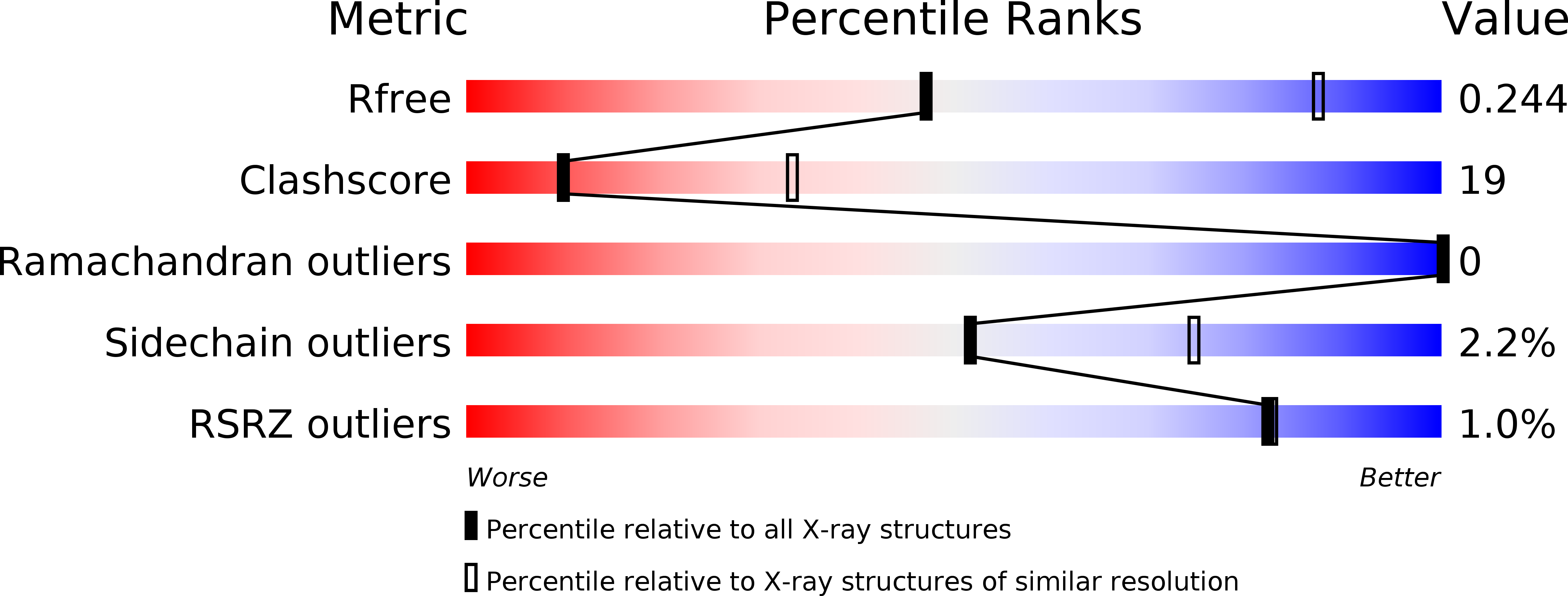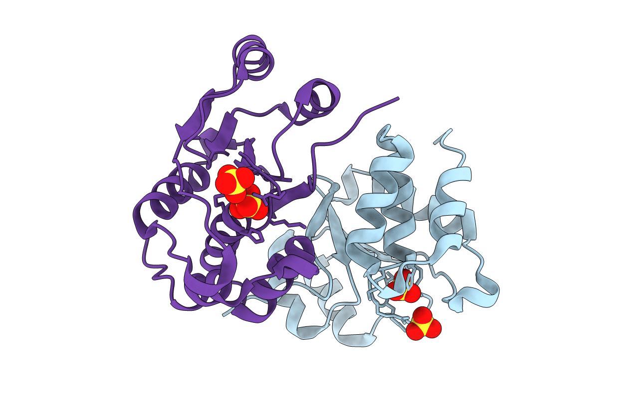
1XRI image
Entry Detail
PDB ID:
1XRI
Title:
X-ray structure of a putative phosphoprotein phosphatase from Arabidopsis thaliana gene AT1G05000
Biological Source:
Source Organism:
Host Organism:
PDB Version:
Deposition Date:
2004-10-14
Release Date:
2004-10-26
Method Details:
Experimental Method:
Resolution:
3.30 Å
R-Value Free:
0.24
R-Value Work:
0.20
Space Group:
P 21 3
Macromolecular Entities
Polymer Type:polypeptide(L)
Description:At1g05000
Chain IDs:A, B
Chain Length:151
Number of Molecules:2
Biological Source:Arabidopsis thaliana
Modified Residue
Compound ID Chain ID Parent Comp ID Details 2D Image
MSE A MET SELENOMETHIONINE
Ligand Molecules
Primary Citation
Structural and functional characterization of a novel phosphatase from the Arabidopsis thaliana gene locus At1g05000.
Proteins 73 241 253 (2008)
PMID: 18433060 DOI: 10.1002/prot.22041

Abstact

The crystal structure of the protein product of the gene locus At1g05000, a hypothetical protein from A. thaliana, was determined by the multiple-wavelength anomalous diffraction method and was refined to an R factor of 20.4% (R(free) = 24.9%) at 3.3 A. The protein adopts the alpha/beta fold found in cysteine phosphatases, a superfamily of phosphatases that possess a catalytic cysteine and form a covalent thiol-phosphate intermediate during the catalytic cycle. In At1g05000, the analogous cysteine (Cys(150)) is located at the bottom of a positively-charged pocket formed by residues that include the conserved arginine (Arg(156)) of the signature active site motif, HCxxGxxRT. Of 74 model phosphatase substrates tested, purified recombinant At1g05000 showed highest activity toward polyphosphate (poly-P(12-13)) and deoxyribo- and ribonucleoside triphosphates, and less activity toward phosphoenolpyruvate, phosphotyrosine, phosphotyrosine-containing peptides, and phosphatidyl inositols. Divalent metal cations were not required for activity and had little effect on the reaction.

Legend

Protein

Chemical

Disease

Primary Citation of related structures