1XRF image
Deposition Date 2004-10-14
Release Date 2005-07-05
Last Version Date 2024-02-14
Entry Detail
PDB ID:
1XRF
Keywords:
Title:
The Crystal Structure of a Novel, Latent Dihydroorotase from Aquifex aeolicus at 1.7 A resolution
Biological Source:
Source Organism:
Host Organism:
Method Details:
Experimental Method:
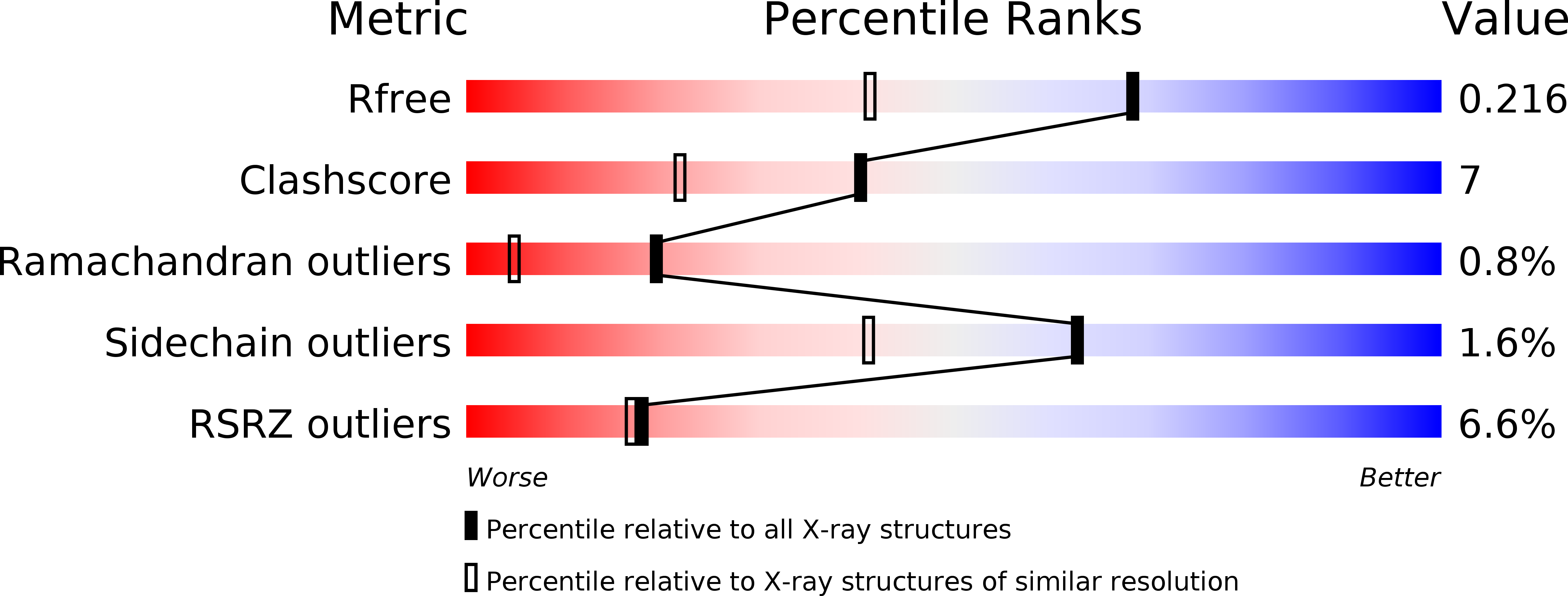
Resolution:
1.65 Å
R-Value Free:
0.21
R-Value Work:
0.17
R-Value Observed:
0.17
Space Group:
C 2 2 21
Macromolecular Entities
Polymer Type:polypeptide(L)
Molecule:Dihydroorotase
Gene (Uniprot):pyrC
Chain IDs:A
Chain Length:467
Number of Molecules:1
Biological Source:Aquifex aeolicus
Primary Citation
The crystal structure of a novel, latent dihydroorotase from Aquifex aeolicus at 1.7A resolution
J.Mol.Biol. 348 535 547 (2005)
PMID: 15826652 DOI: 10.1016/j.jmb.2005.03.015

Abstact

Dihydroorotases (EC 3.5.2.3) catalyze the reversible cyclization of carbamoyl aspartate to form dihydroorotate in de novo pyrimidine biosynthesis. The X-ray structures of Aquifex aeolicus dihydroorotase in two space groups, C222(1) and C2, were determined at a resolution of 1.7A. These are the first structures of a type I dihydroorotase, a class of molecules that includes the dihydroorotase domain of mammalian CAD. The type I enzymes are more ancient and larger, at 45 kDa, than the type II enzymes exemplified by the 38 kDa Escherichia coli dihydroorotase. Both dihydroorotases are members of the metallo-dependent hydrolase superfamily, whose members have a distorted "TIM barrel" domain containing the active site. However, A.aeolicus dihydroorotase has a second, composite domain, which the E.coli enzyme lacks and has only one of the two zinc atoms present in the E.coli enzyme. A.aeolicus dihydroorotase is unique in exhibiting significant activity only when complexed with aspartate transcarbamoylase, whereas the E.coli dihydroorotase and the CAD dihydroorotase domain are active as free proteins. The latency of A.aeolicus dihydroorotase can be related to two differences between its structure and that of E.coli dihydroorotase: (1) the monoclinic structure has a novel cysteine ligand to the zinc that blocks the active site and possibly functions as a "cysteine switch"; and (2) active site residues that bind the substrate in E.coli dihydroorotase are located in disordered loops in both crystal structures of A.aeolicus dihydroorotase and may function as a disorder-to-order "entropy switch".

Legend

Protein

Chemical

Disease

Primary Citation of related structures