1XRE image
Deposition Date 2004-10-14
Release Date 2005-07-19
Last Version Date 2023-08-23
Entry Detail
PDB ID:
1XRE
Keywords:
Title:
Crystal Structure of SodA-2 (BA5696) from Bacillus anthracis at 1.8A Resolution.
Biological Source:
Source Organism:
Host Organism:
Method Details:
Experimental Method:
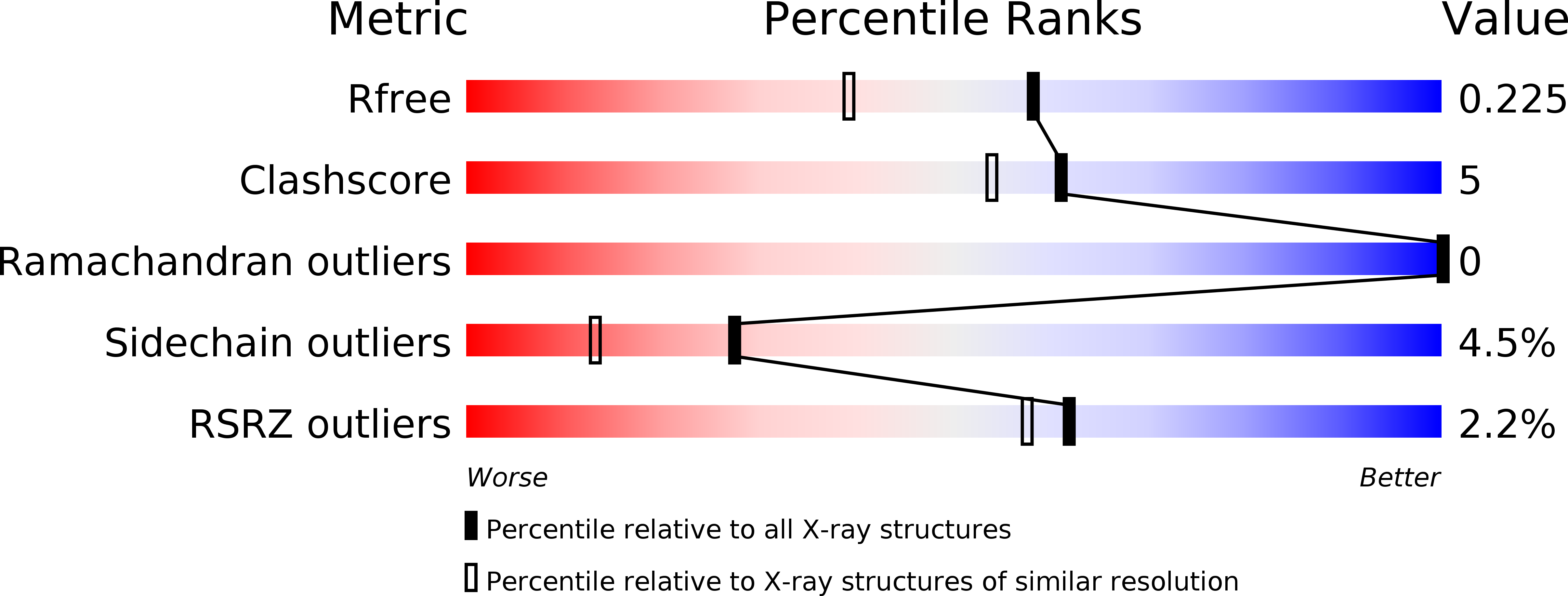
Resolution:
1.80 Å
R-Value Free:
0.21
R-Value Work:
0.15
R-Value Observed:
0.16
Space Group:
P 1 21 1
Macromolecular Entities
Polymer Type:polypeptide(L)
Molecule:Superoxide dismutase
Gene (Uniprot):sodA2
Chain IDs:A, B
Chain Length:217
Number of Molecules:2
Biological Source:Bacillus anthracis
Ligand Molecules
Primary Citation
Structures of two superoxide dismutases from Bacillus anthracis reveal a novel active centre.
Acta Crystallogr.,Sect.F 61 621 624 (2005)
PMID: 16511113 DOI: 10.1107/S1744309105017380

Abstact

The BA4499 and BA5696 genes of Bacillus anthracis encode proteins homologous to manganese superoxide dismutase, suggesting that this organism has an expanded repertoire of antioxidant proteins. Differences in metal specificity and quaternary structure between the dismutases of prokaryotes and higher eukaryotes may be exploited in the development of therapeutic antibacterial compounds. Here, the crystal structure of two Mn superoxide dismutases from B. anthracis solved to high resolution are reported. Comparison of their structures reveals that a highly conserved residue near the active centre is substituted in one of the proteins and that this is a characteristic feature of superoxide dismutases from the B. cereus/B. anthracis/B. thuringiensis group of organisms.

Legend

Protein

Chemical

Disease

Primary Citation of related structures