1XQS image
Deposition Date 2004-10-13
Release Date 2005-03-01
Last Version Date 2024-05-29
Entry Detail
PDB ID:
1XQS
Keywords:
Title:
Crystal structure of the HspBP1 core domain complexed with the fragment of Hsp70 ATPase domain
Biological Source:
Source Organism:
Homo sapiens (Taxon ID: 9606)
Host Organism:
Method Details:
Experimental Method:
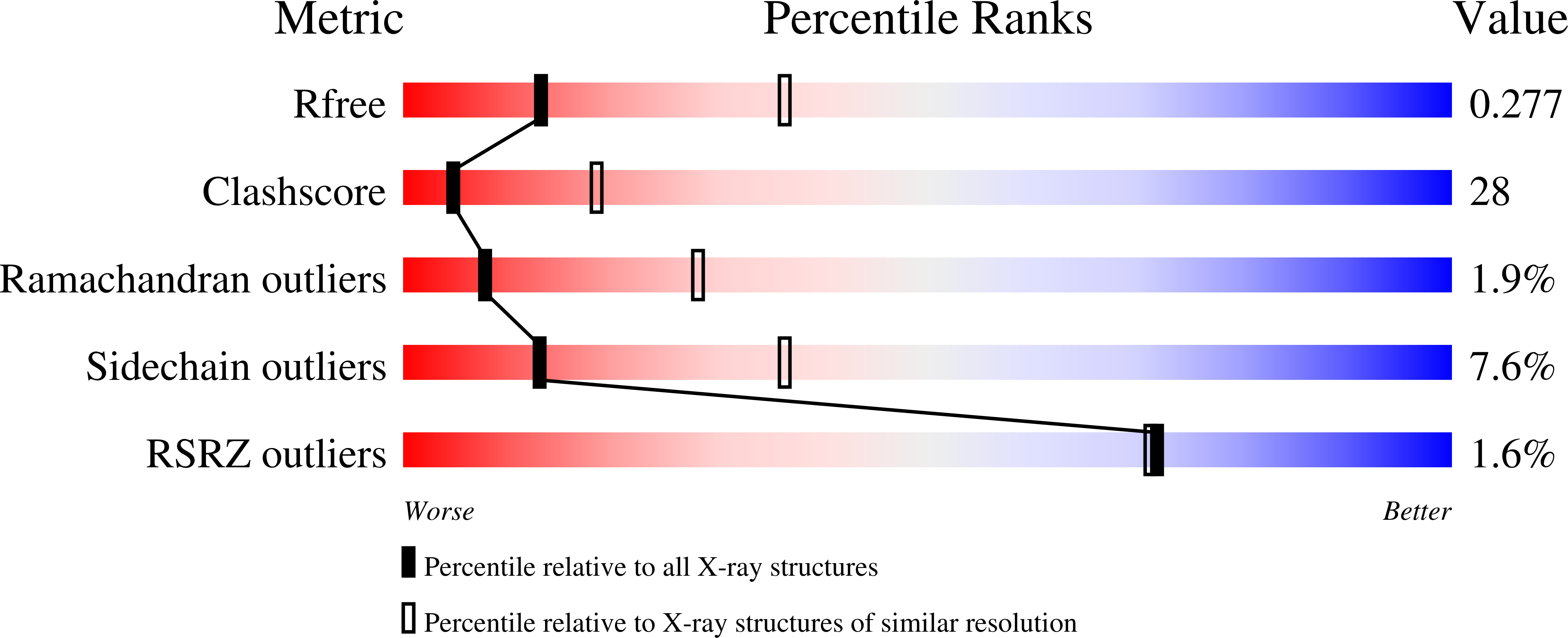
Resolution:
2.90 Å
R-Value Free:
0.29
R-Value Work:
0.23
Space Group:
P 21 21 21
Macromolecular Entities
Polymer Type:polypeptide(L)
Molecule:HSPBP1 protein
Gene (Uniprot):HSPBP1
Mutations:E88G
Chain IDs:A, B
Chain Length:280
Number of Molecules:2
Biological Source:Homo sapiens
Polymer Type:polypeptide(L)
Molecule:Heat shock 70 kDa protein 1
Chain IDs:C, D
Chain Length:191
Number of Molecules:2
Biological Source:Homo sapiens
Ligand Molecules
Primary Citation
Regulation of Hsp70 Function by HspBP1; Structural Analysis Reveals an Alternate Mechanism for Hsp70 Nucleotide Exchange
Mol.Cell 17 367 379 (2005)
PMID: 15694338 DOI: 10.1016/j.molcel.2004.12.023

Abstact

HspBP1 belongs to a family of eukaryotic proteins recently identified as nucleotide exchange factors for Hsp70. We show that the S. cerevisiae ortholog of HspBP1, Fes1p, is required for efficient protein folding in the cytosol at 37 degrees C. The crystal structure of HspBP1, alone and complexed with part of the Hsp70 ATPase domain, reveals a mechanism for its function distinct from that of BAG-1 or GrpE, previously characterized nucleotide exchange factors of Hsp70. HspBP1 has a curved, all alpha-helical fold containing four armadillo-like repeats unlike the other nucleotide exchange factors. The concave face of HspBP1 embraces lobe II of the ATPase domain, and a steric conflict displaces lobe I, reducing the affinity for nucleotide. In contrast, BAG-1 and GrpE trigger a conserved conformational change in lobe II of the ATPase domain. Thus, nucleotide exchange on eukaryotic Hsp70 occurs through two distinct mechanisms.

Legend

Protein

Chemical

Disease

Primary Citation of related structures