Deposition Date
2004-10-08
Release Date
2004-11-09
Last Version Date
2024-11-13
Entry Detail
PDB ID:
1XP4
Keywords:
Title:
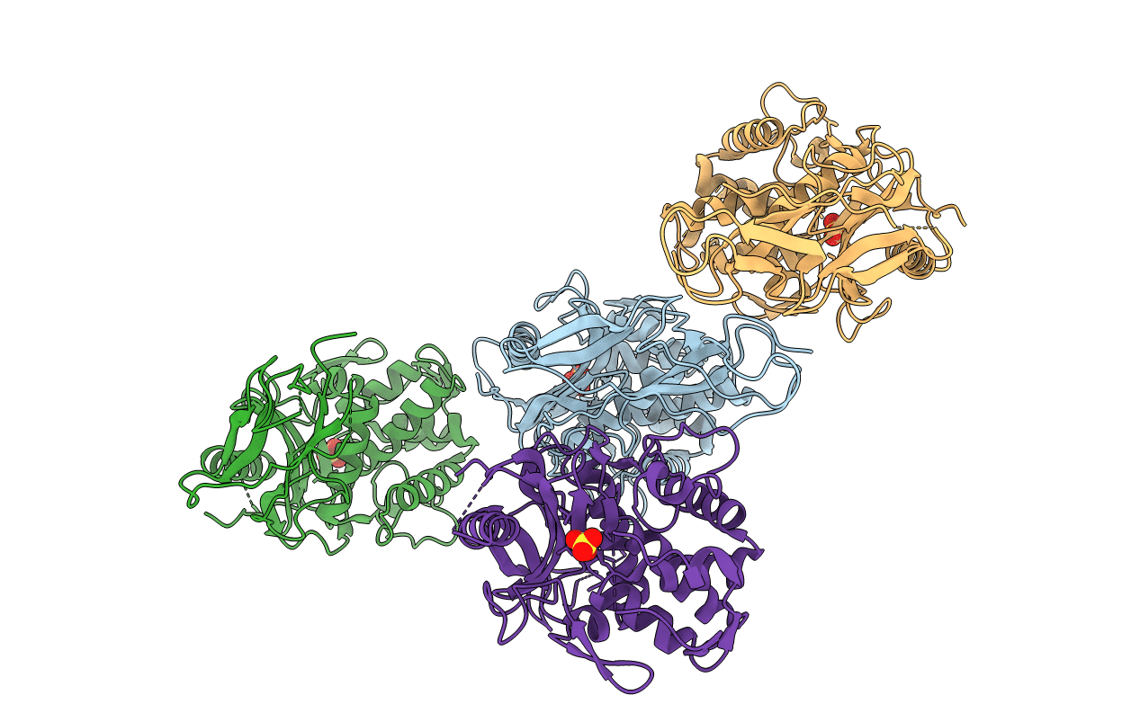
Crystal structure of a peptidoglycan synthesis regulatory factor (PBP3) from Streptococcus pneumoniae
Biological Source:
Source Organism(s):
Streptococcus pneumoniae R6 (Taxon ID: 171101)
Expression System(s):
Method Details:
Experimental Method:
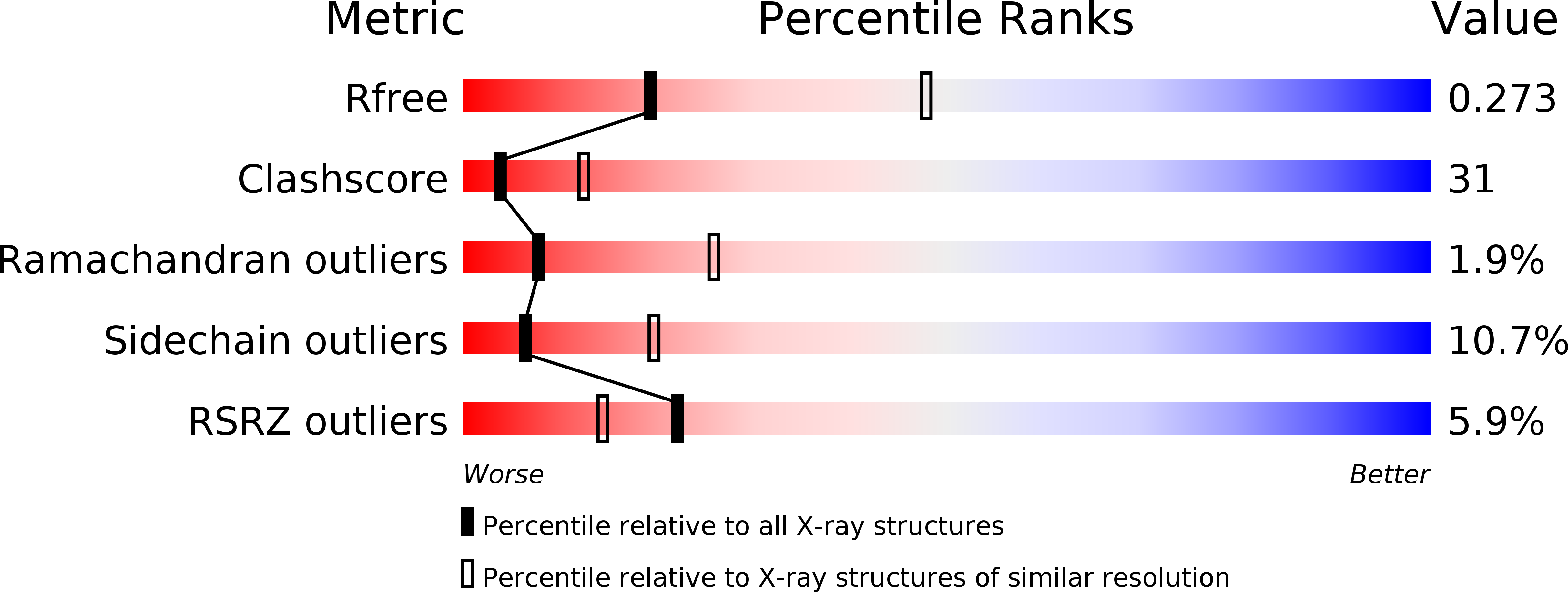
Resolution:
2.80 Å
R-Value Free:
0.27
R-Value Work:
0.23
R-Value Observed:
0.23
Space Group:
P 21 21 21