1XOP image
Deposition Date 2004-10-06
Release Date 2005-09-27
Last Version Date 2024-05-22
Entry Detail
PDB ID:
1XOP
Keywords:
Title:
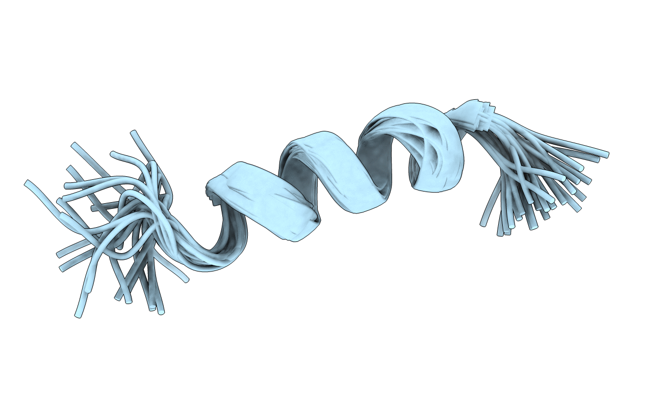
NMR structure of G1V mutant of influenza hemagglutinin fusion peptide in DPC micelles at pH 5
Biological Source:
Source Organism:
Method Details:
Experimental Method:
Conformers Calculated:
25
Conformers Submitted:
25
Selection Criteria:
structures with the lowest energy
Macromolecular Entities
Structures with similar UniProt ID
Protein Blast
Polymer Type:polypeptide(L)
Molecule:Hemagglutinin
Chain IDs:A
Chain Length:20
Number of Molecules:1
Biological Source:
Ligand Molecules
Primary Citation
Membrane structures of the hemifusion-inducing fusion peptide mutant G1S and the fusion-blocking mutant G1V of influenza virus hemagglutinin suggest a mechanism for pore opening in membrane fusion.
J.Virol. 79 12065 12076 (2005)
PMID: 16140782 DOI: 10.1128/JVI.79.18.12065-12076.2005

Abstact

Influenza virus hemagglutinin (HA)-mediated membrane fusion is initiated by a conformational change that releases a V-shaped hydrophobic fusion domain, the fusion peptide, into the lipid bilayer of the target membrane. The most N-terminal residue of this domain, a glycine, is highly conserved and is particularly critical for HA function; G1S and G1V mutant HAs cause hemifusion and abolish fusion, respectively. We have determined the atomic resolution structures of the G1S and G1V mutant fusion domains in membrane environments. G1S forms a V with a disrupted "glycine edge" on its N-terminal arm and G1V adopts a slightly tilted linear helical structure in membranes. Abolishment of the kink in G1V results in reduced hydrophobic penetration of the lipid bilayer and an increased propensity to form beta-structures at the membrane surface. These results underline the functional importance of the kink in the fusion peptide and suggest a structural role for the N-terminal glycine ridge in viral membrane fusion.

Legend

Protein

Chemical

Disease

Primary Citation of related structures