Deposition Date
2004-10-05
Release Date
2004-12-07
Last Version Date
2024-02-14
Entry Detail
PDB ID:
1XO5
Keywords:
Title:
Crystal structure of CIB1, an EF-hand, integrin and kinase-binding protein
Biological Source:
Source Organism:
Homo sapiens (Taxon ID: 9606)
Host Organism:
Method Details:
Experimental Method:
Resolution:
1.99 Å
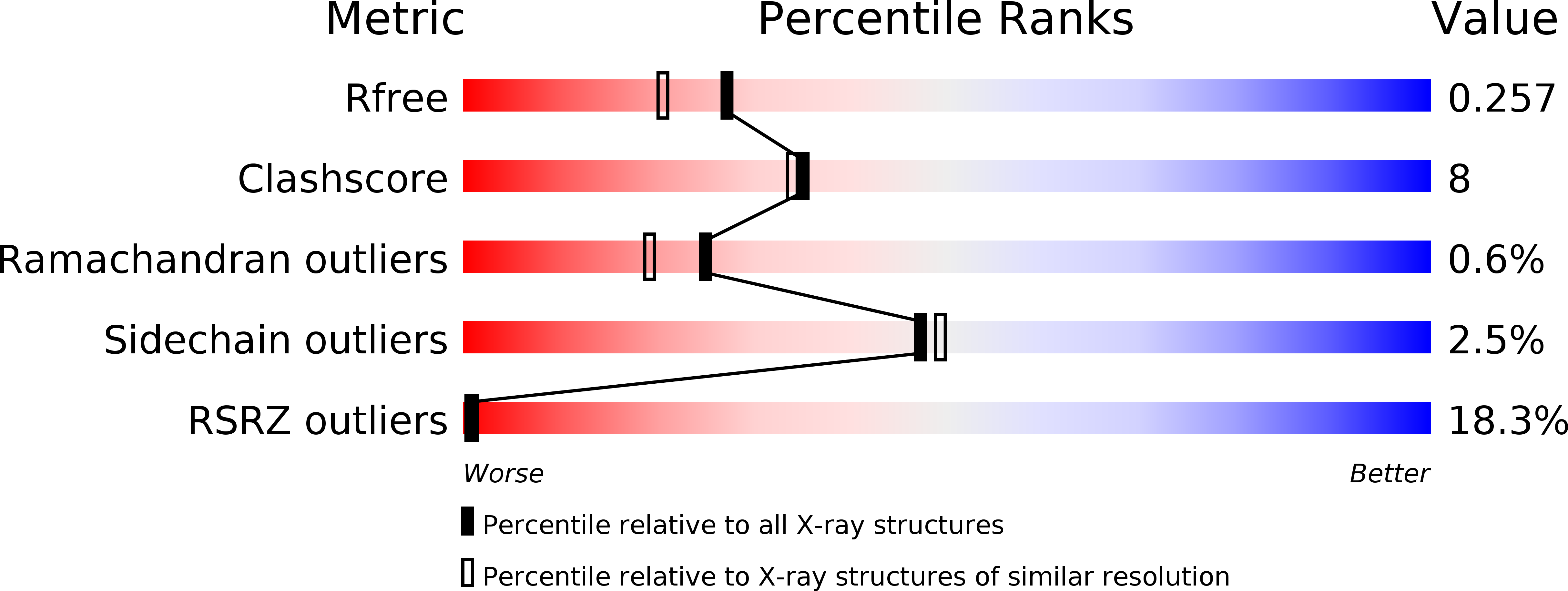
R-Value Free:
0.25
R-Value Work:
0.21
R-Value Observed:
0.21
Space Group:
P 1 21 1