Deposition Date
2004-10-04
Release Date
2005-03-22
Last Version Date
2024-10-30
Entry Detail
PDB ID:
1XN3
Keywords:
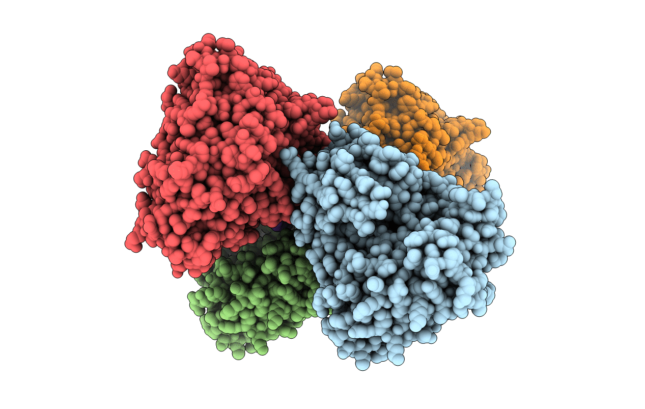
Title:
Crystal structure of Beta-secretase bound to a long inhibitor with additional upstream residues.
Biological Source:
Source Organism:
Homo sapiens (Taxon ID: 9606)
Host Organism:
Method Details:
Experimental Method:
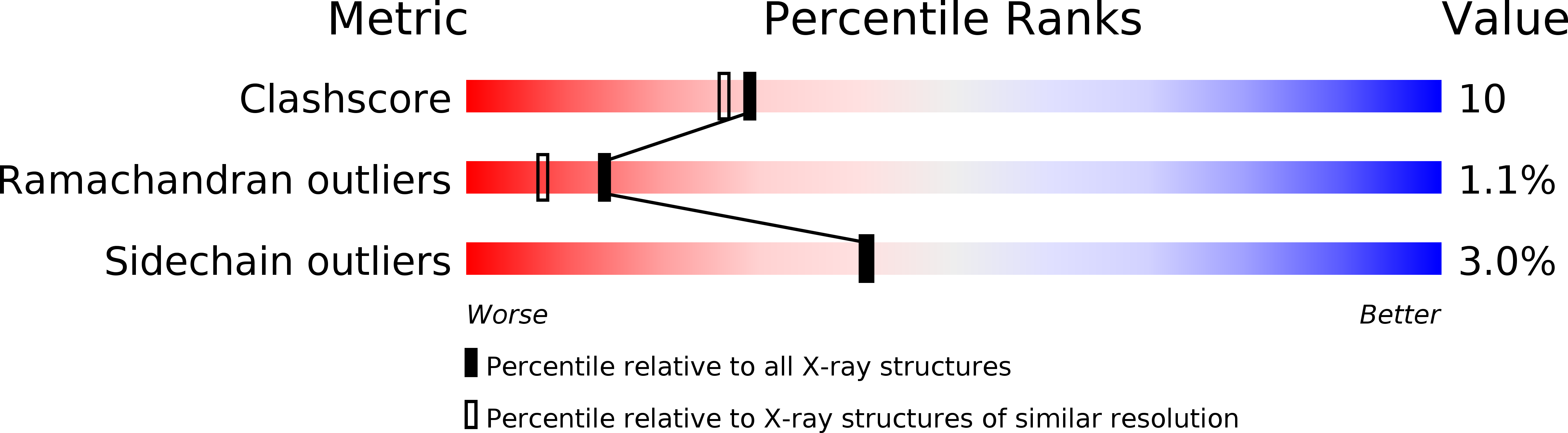
Resolution:
2.00 Å
R-Value Free:
0.23
R-Value Observed:
0.20
Space Group:
P 1 21 1