Deposition Date
2004-10-01
Release Date
2004-10-12
Last Version Date
2024-02-14
Entry Detail
PDB ID:
1XM5
Keywords:
Title:
Crystal structure of metal-dependent hydrolase ybeY from E. coli, Pfam UPF0054
Biological Source:
Source Organism(s):
Escherichia coli (Taxon ID: 562)
Expression System(s):
Method Details:
Experimental Method:
Resolution:
2.70 Å
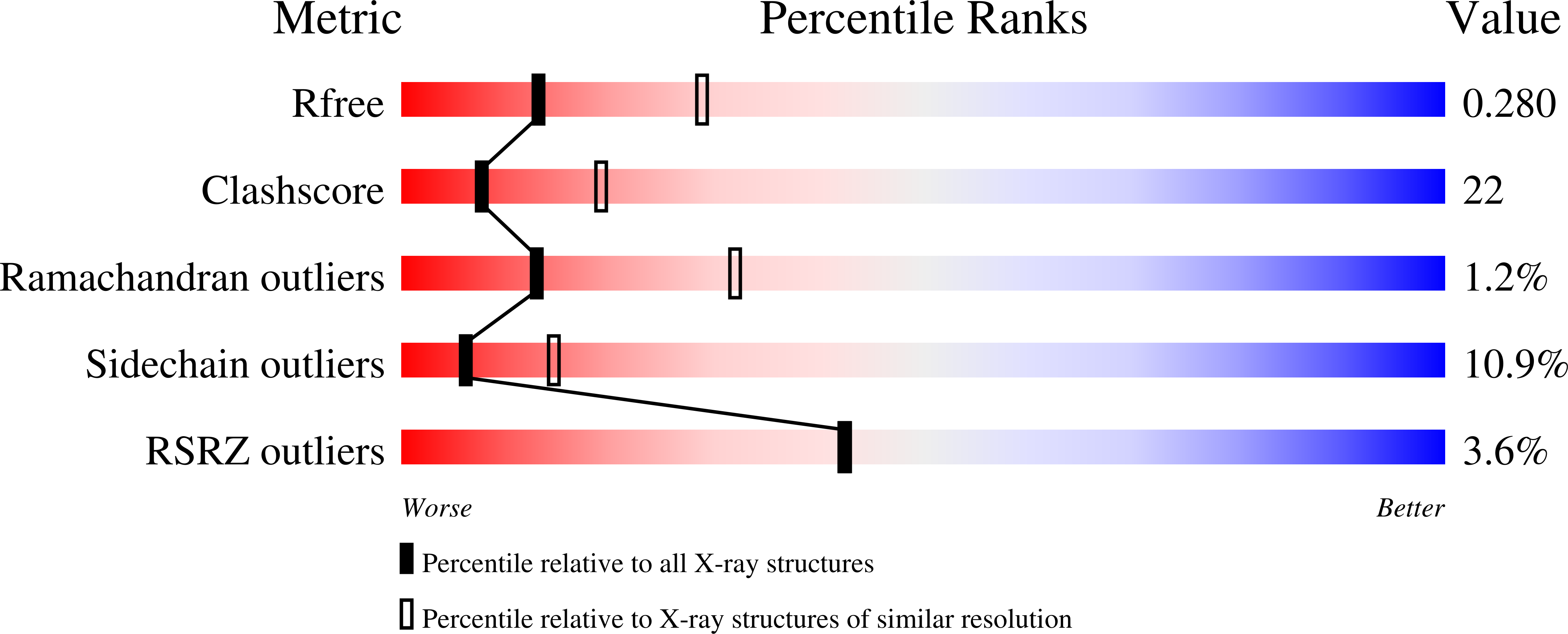
R-Value Free:
0.27
R-Value Work:
0.23
R-Value Observed:
0.23
Space Group:
P 21 21 21