1XKI image
Deposition Date 2004-09-29
Release Date 2004-10-19
Last Version Date 2024-05-29
Entry Detail
PDB ID:
1XKI
Title:
Crystal structure of human tear lipocalin/von Ebners gland protein
Biological Source:
Source Organism:
Homo sapiens (Taxon ID: 9606)
Host Organism:
Method Details:
Experimental Method:
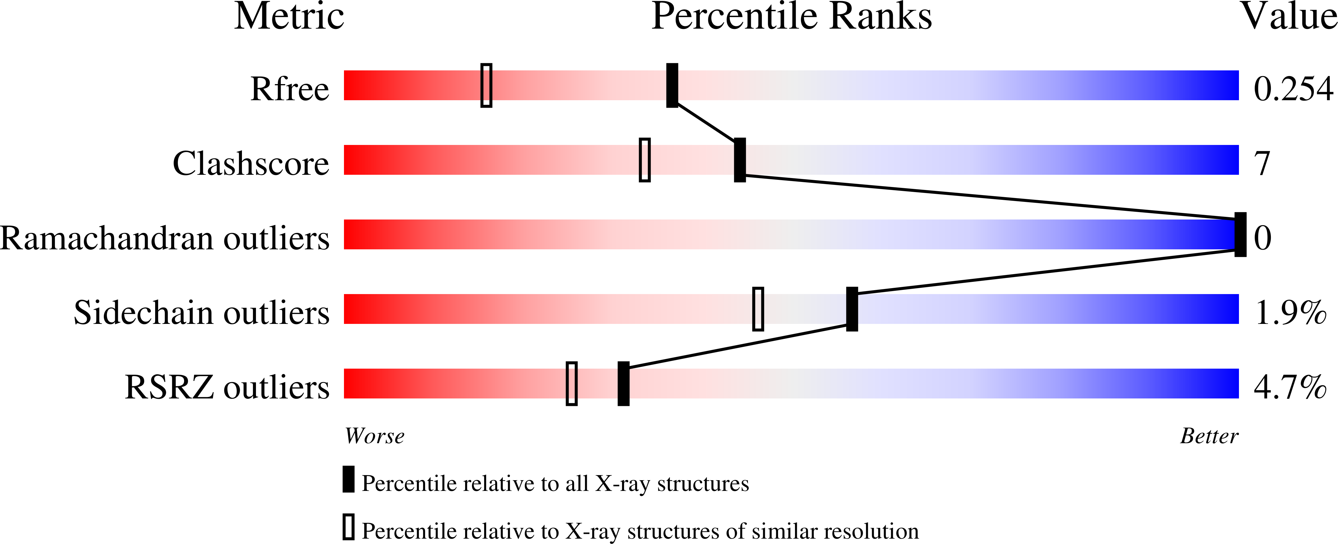
Resolution:
1.80 Å
R-Value Free:
0.25
R-Value Work:
0.18
R-Value Observed:
0.19
Space Group:
C 1 2 1
Macromolecular Entities
Structures with similar UniProt ID
Protein Blast
Polymer Type:polypeptide(L)
Molecule:Von Ebner's gland protein
Gene (Uniprot):LCN1
Mutagens:C101S, D158A
Chain IDs:A
Chain Length:162
Number of Molecules:1
Biological Source:Homo sapiens
Primary Citation
The 1.8-A crystal structure of human tear lipocalin reveals an extended branched cavity with capacity for multiple ligands
J.Biol.Chem. 280 484 493 (2005)
PMID: 15489503 DOI: 10.1074/jbc.M410466200

Abstact

In contrast with earlier assumptions, which classified human tear lipocalin (Tlc) as an outlier member of the lipocalin protein family, the 1.8-A resolution crystal structure of the recombinant apoprotein confirms the typical eight-stranded antiparallel beta-barrel architecture with an alpha-helix attached to it. The fold of Tlc most closely resembles the bovine dander allergen Bos d 2, a well characterized prototypic lipocalin, but also reveals similarity with beta-lactoglobulin. However, compared with other lipocalin structures Tlc exhibits an extremely wide ligand pocket, whose entrance is formed by four partially disordered loops. The cavity deeply extends into the beta-barrel structure, where it ends in two distinct lobes. This unusual structural feature explains the known promiscuity of Tlc for various ligands, with chemical structures ranging from lipids and retinoids to the macrocyclic antibiotic rifampin and even to microbial siderophores. Notably, earlier findings of biological activity as a thiol protease inhibitor have no correspondence in the three-dimensional structure of Tlc, rather it appears that its proteolytic fragments could be responsible for this phenomenon. Hence, the present structural analysis sheds new light on the ligand binding activity of this functionally obscure but abundant human lipocalin.

Legend

Protein

Chemical

Disease

Primary Citation of related structures
Feedback Form
Name
Email
Institute
Feedback