1XJX image
Deposition Date 2004-09-26
Release Date 2005-03-15
Last Version Date 2024-04-03
Entry Detail
PDB ID:
1XJX
Keywords:
Title:
The crystal structures of the DNA binding sites of the RUNX1 transcription factor
Biological Source:
Source Organism:
(Taxon ID: ) (Taxon ID: )
Method Details:
Experimental Method:
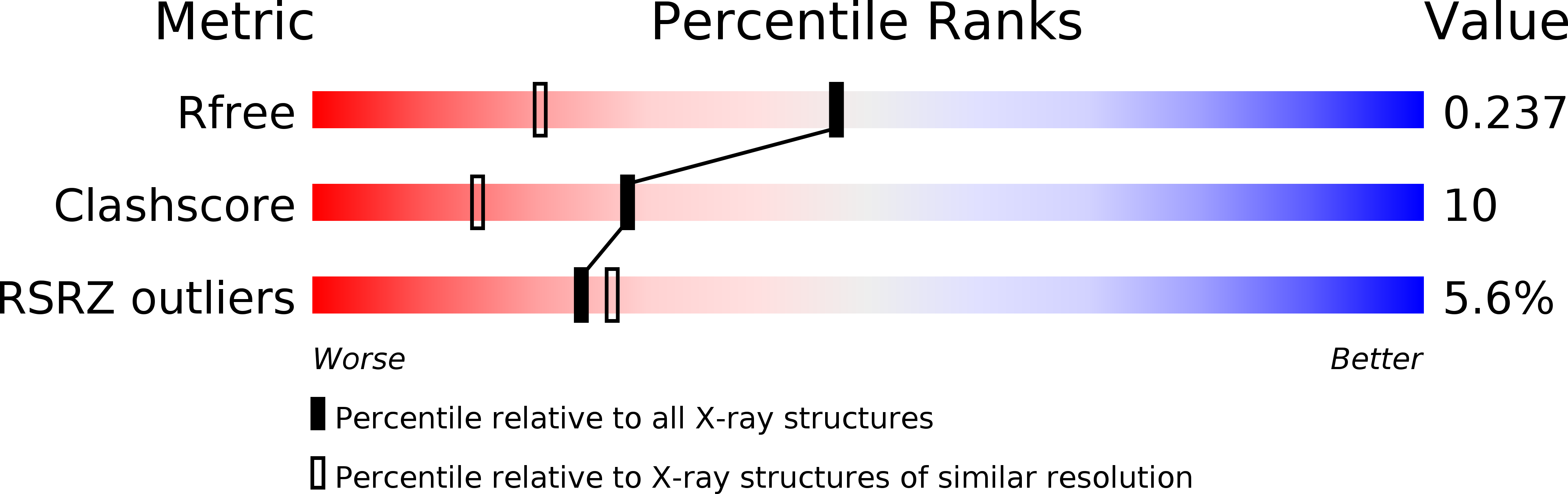
Resolution:
1.70 Å
R-Value Free:
0.25
R-Value Work:
0.16
R-Value Observed:
0.16
Space Group:
P 43
Macromolecular Entities
Polymer Type:polydeoxyribonucleotide
Molecule:5'-D(*TP*CP*TP*GP*CP*GP*GP*TP*C)-3'
Chain IDs:A
Chain Length:9
Number of Molecules:1
Biological Source:
Polymer Type:polydeoxyribonucleotide
Molecule:5'-D(*TP*GP*AP*CP*CP*GP*CP*AP*G)-3'
Chain IDs:B
Chain Length:9
Number of Molecules:1
Biological Source:
Primary Citation
Structures of the DNA-binding site of Runt-domain transcription regulators.
Acta Crystallogr.,Sect.D 61 236 246 (2005)
PMID: 15735333 DOI: 10.1107/S0907444904032378

Abstact

Runt-domain (RD) proteins are transcription factors that play fundamental roles in various developmental pathways. They bind specifically to DNA sequences of the general form PyGPyGGTPy (Py = pyrimidine), through which they regulate transcription of target genes. The DNA duplex TCTGCGGTC/TGACCGCAG, incorporating the binding site for the RD transcription factors (bold), was crystallized in space group P4(3). X-ray analysis of two crystals diffracting to 1.7 and 2.0 angstroms resolution, which had slight variations in their unit-cell parameters, revealed two distinct conformations of the A-DNA helix. The two crystal structures possessed several structure and hydration features that had previously been observed in A-DNA duplexes. A comparative analysis of the present A-DNA structures and those of previously reported B-DNA crystal structures of RD-binding sites in free and protein-bound states showed the various duplexes to display several common features. Within this series, the present A-DNA duplexes adopt two conformations along the pathway from the canonical A-DNA to the B-DNA forms and the protein-bound helices display conformational features that are intermediate between those of the current A-DNA structures and that of the B-DNA-type helix of the free RD target. Based on these data and energy considerations, it is likely that the propensity of the RD-binding site to adopt the A-DNA or B-DNA conformation in solution depends on the sequence context and environmental conditions, and that the transition from either DNA form to the protein-bound conformation involves a small energy barrier.

Legend

Protein

Chemical

Disease

Primary Citation of related structures