Deposition Date
2004-09-21
Release Date
2004-12-07
Last Version Date
2024-02-14
Entry Detail
PDB ID:
1XI1
Keywords:
Title:
Phi29 DNA polymerase ssDNA complex, monoclinic crystal form
Biological Source:
Source Organism(s):
Bacillus phage phi29 (Taxon ID: 10756)
Expression System(s):
Method Details:
Experimental Method:
Resolution:
2.20 Å
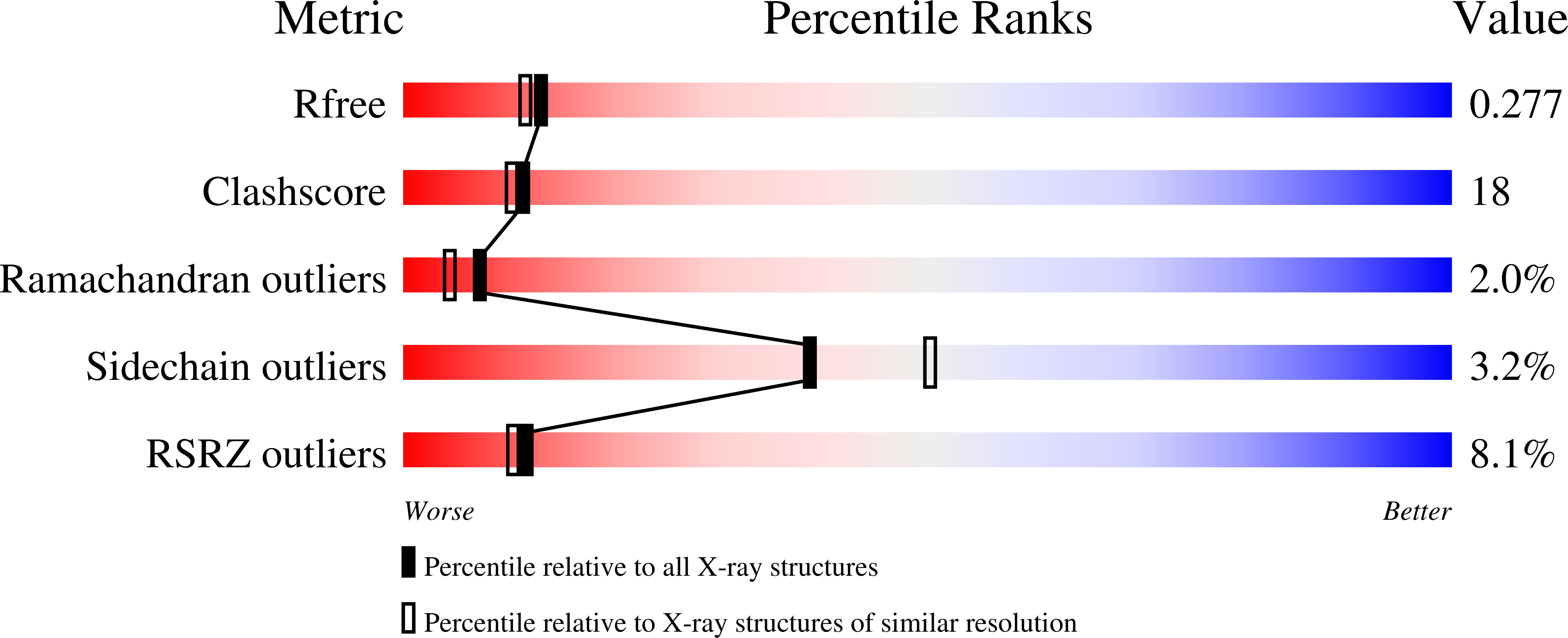
R-Value Free:
0.27
R-Value Work:
0.24
R-Value Observed:
0.24
Space Group:
P 1 21 1