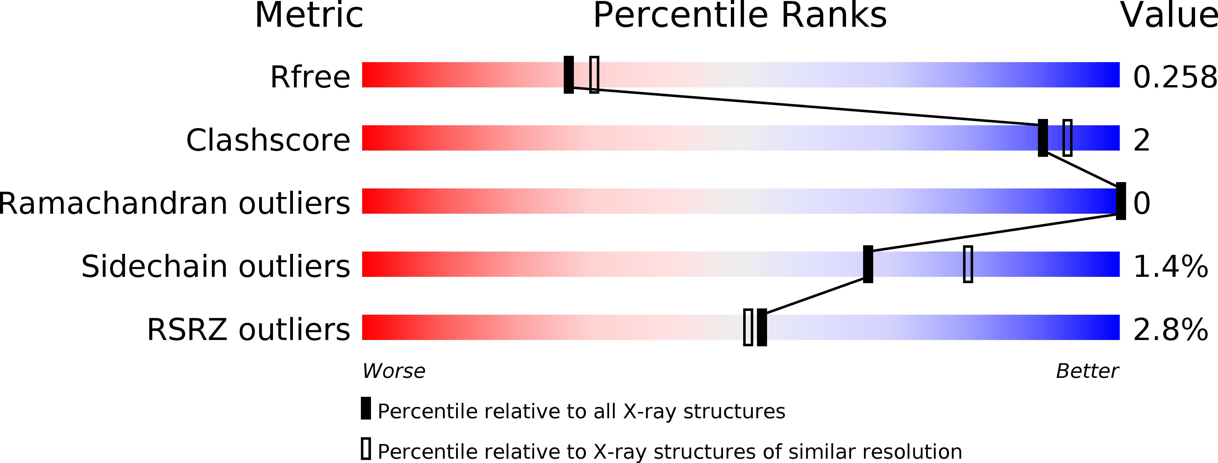
1XHO image
Deposition Date 2004-09-20
Release Date 2004-11-23
Last Version Date 2024-11-13
Entry Detail
PDB ID:
1XHO
Title:
Chorismate mutase from Clostridium thermocellum Cth-682
Biological Source:
Source Organism:
Host Organism:
Method Details:
Experimental Method:
Resolution:
2.20 Å
R-Value Free:
0.25
R-Value Work:
0.19
R-Value Observed:
0.19
Space Group:
P 31
Macromolecular Entities
Structures with similar UniProt ID
Protein Blast
Polymer Type:polypeptide(L)
Molecule:Chorismate mutase
Chain IDs:A, B, C
Chain Length:148
Number of Molecules:3
Biological Source:Clostridium thermocellum
Modified Residue
Compound ID Chain ID Parent Comp ID Details 2D Image
MSE A MET SELENOMETHIONINE
Ligand Molecules
Primary Citation
Away from the edge II: in-house Se-SAS phasing with chromium radiation.
Acta Crystallogr.,Sect.D 61 960 966 (2005)
PMID: 15983419 DOI: 10.1107/S0907444905010644

Abstact

Recently, the demands of high-throughput macromolecular crystallography have driven continuous improvements in phasing methods, data-collection protocols and many other technologies. Single-wavelength anomalous scattering (SAS) phasing with chromium X-ray radiation opens a new possibility for phasing a protein with data collected in-house and has led to several successful examples of de novo structure solution using only weak anomalous scatterers such as sulfur. To further reduce data-collection time and make SAS phasing more robust, it is natural to combine selenomethionine-derivatized protein (SeMet protein) with Cr Kalpha radiation to take advantage of the larger anomalous scattering signal from selenium (f'' = 2.28 e(-)) compared with sulfur (f'' = 1.14 e(-)). As reported herein, the crystal structure of a putative chorismate mutase from Clostridium thermocellum was determined using Se-SAS with Cr Kalpha radiation. Each protein molecule contains eight selenomethionine residues in 148 amino-acid residues, providing a calculated Bijvoet ratio of about 3.5% at the Cr Kalpha wavelength. A single data set to 2.2 A resolution with approximately ninefold redundancy was collected using an imaging-plate detector coupled with a Cr source. Structure solution, refinement and deposition to the Protein Data Bank were performed within 9 h of the availability of the scaled diffraction data. The procedure used here is applicable to many other proteins and promises to become a routine pathway for in-house high-throughput crystallography.

Legend

Protein

Chemical

Disease

Primary Citation of related structures