1XHM image
Deposition Date 2004-09-20
Release Date 2005-08-09
Last Version Date 2023-08-23
Entry Detail
PDB ID:
1XHM
Title:
The Crystal Structure of a Biologically Active Peptide (SIGK) Bound to a G Protein Beta:Gamma Heterodimer
Biological Source:
Source Organism:
Bos taurus (Taxon ID: 9913)
Host Organism:
Method Details:
Experimental Method:
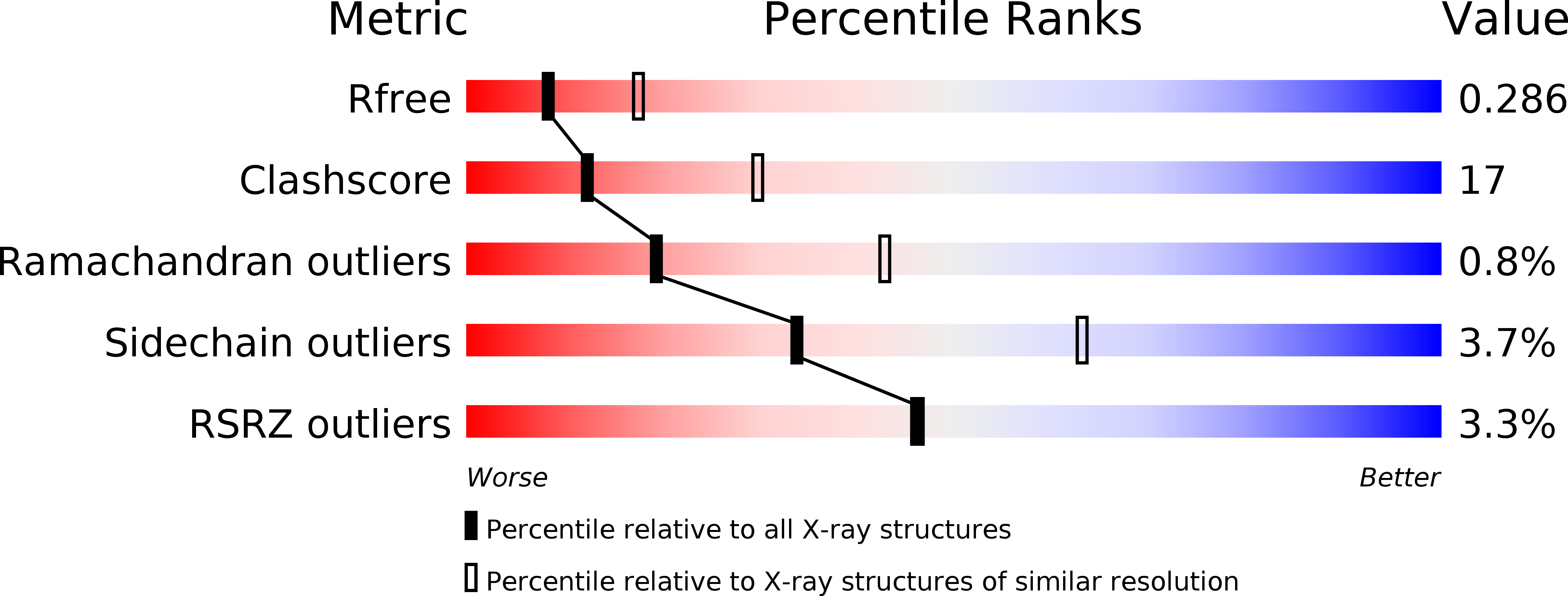
Resolution:
2.70 Å
R-Value Free:
0.28
R-Value Work:
0.22
R-Value Observed:
0.22
Space Group:
P 21 21 21
Macromolecular Entities
Structures with similar UniProt ID
Protein Blast
Polymer Type:polypeptide(L)
Molecule:Guanine nucleotide-binding protein G(I)/G(S)/G(T) beta subunit 1
Gene (Uniprot):GNB1
Chain IDs:A
Chain Length:340
Number of Molecules:1
Biological Source:Bos taurus
Structures with similar UniProt ID
Protein Blast
Polymer Type:polypeptide(L)
Molecule:Guanine nucleotide-binding protein G(I)/G(S)/G(O) gamma-2 subunit
Gene (Uniprot):GNG2
Chain IDs:B
Chain Length:77
Number of Molecules:1
Biological Source:Bos taurus
Polymer Type:polypeptide(L)
Molecule:SIGK Peptide
Chain IDs:C
Chain Length:15
Number of Molecules:1
Biological Source:
Primary Citation
Structural and Molecular Characterization of a Preferred Protein Interaction Surface on G Protein betagamma Subunits.
Biochemistry 44 10593 10604 (2005)
PMID: 16060668 DOI: 10.1021/bi050655i

Abstact

G protein betagamma subunits associate with many binding partners in cellular signaling cascades. In previous work, we used random-peptide phage display screening to identify a diverse family of peptides that bound to a common surface on Gbetagamma subunits and blocked a subset of Gbetagamma effectors. Later studies showed that one of the peptides caused G protein activation through a novel Gbetagamma-dependent, nucleotide exchange-independent mechanism. Here we report the X-ray crystal structure of Gbeta(1)gamma(2) bound to this peptide, SIGK (SIGKAFKILGYPDYD), at 2.7 A resolution. SIGK forms a helical structure that binds the same face of Gbeta(1) as the switch II region of Galpha. The interaction interface can be subdivided into polar and nonpolar interfaces that together contain a mixture of binding determinants that may be responsible for the ability of this surface to recognize multiple protein partners. Systematic mutagenic analysis of the peptide-Gbeta(1) interface indicates that distinct sets of amino acids within this interface are required for binding of different peptides. Among these unique amino acid interactions, specific electrostatic binding contacts within the polar interface are required for peptide-mediated subunit dissociation. The data provide a mechanistic basis for multiple target recognition by Gbetagamma subunits with diverse functional interactions within a common interface and suggest that pharmacological targeting of distinct regions within this interface could allow for selective manipulation of Gbetagamma-dependent signaling pathways.

Legend

Protein

Chemical

Disease

Primary Citation of related structures
Feedback Form
Name
Email
Institute
Feedback