Deposition Date
2004-09-17
Release Date
2004-10-26
Last Version Date
2024-11-13
Entry Detail
PDB ID:
1XHB
Keywords:
Title:
The Crystal Structure of UDP-GalNAc: polypeptide alpha-N-acetylgalactosaminyltransferase-T1
Biological Source:
Source Organism(s):
Mus musculus (Taxon ID: 10090)
Expression System(s):
Method Details:
Experimental Method:
Resolution:
2.50 Å
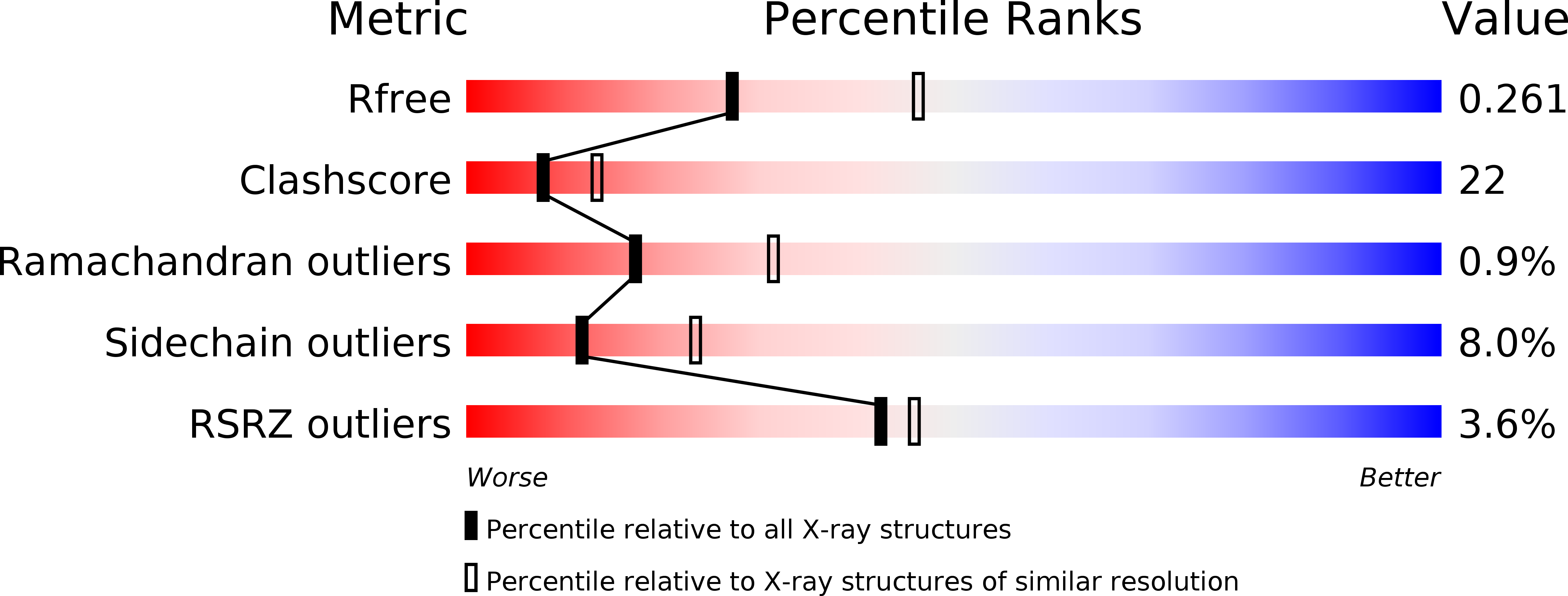
R-Value Free:
0.25
R-Value Work:
0.21
R-Value Observed:
0.21
Space Group:
P 43