1XGE image
Deposition Date 2004-09-17
Release Date 2005-04-26
Last Version Date 2023-11-15
Entry Detail
PDB ID:
1XGE
Keywords:
Title:
Dihydroorotase from Escherichia coli: Loop Movement and Cooperativity between subunits
Biological Source:
Source Organism:
Escherichia coli (Taxon ID: 562)
Host Organism:
Method Details:
Experimental Method:
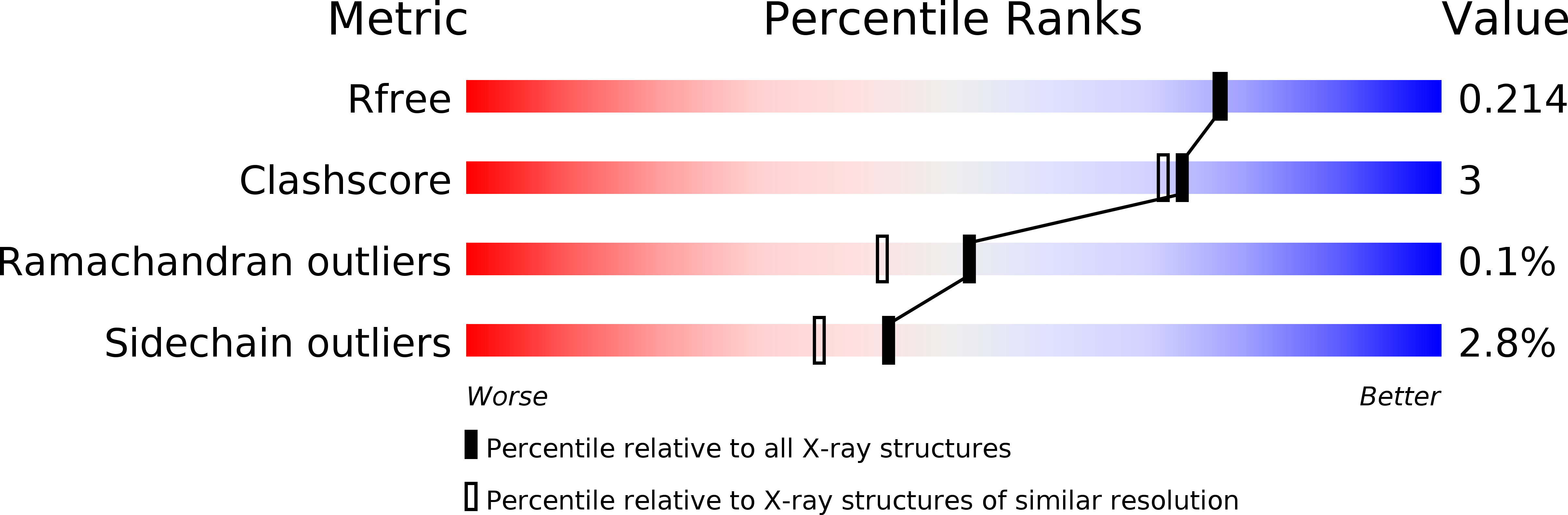
Resolution:
1.90 Å
R-Value Free:
0.21
R-Value Work:
0.16
R-Value Observed:
0.17
Space Group:
P 21 21 21
Macromolecular Entities
Structures with similar UniProt ID
Protein Blast
Polymer Type:polypeptide(L)
Molecule:Dihydroorotase
Gene (Uniprot):pyrC
Chain IDs:A, B
Chain Length:347
Number of Molecules:2
Biological Source:Escherichia coli
Modified Residue
Compound ID Chain ID Parent Comp ID Details 2D Image
KCX A LYS LYSINE NZ-CARBOXYLIC ACID
Primary Citation
Dihydroorotase from Escherichia coli: Loop Movement and Cooperativity between Subunits
J.Mol.Biol. 348 523 533 (2005)
PMID: 15826651 DOI: 10.1016/j.jmb.2005.01.067

Abstact

Escherichia coli dihydroorotase has been crystallized in the presence of the product, L-dihydroorotate (L-DHO), and the structure refined at 1.9A resolution. The structure confirms that previously reported (PDB entry 1J79), crystallized in the presence of the substrate N-carbamyl-D,L-aspartate (D, L-CA-asp), which had a dimer in the asymmetric unit, with one subunit having the substrate, L-CA-asp bound at the active site and the other having L-DHO. Importantly, no explanation for the unusual structure was given. Our results now show that a loop comprised of residues 105-115 has different conformations in the two subunits. In the case of the L-CA-asp-bound subunit, this loop reaches in toward the active site and makes hydrogen-bonding contact with the bound substrate molecule. For the L-DHO-bound subunit, the loop faces in the opposite direction and forms part of the surface of the protein. Analysis of the kinetics for conversion of L-DHO to L-CA-asp at low concentrations of L-DHO shows positive cooperativity with a Hill coefficient n=1.57(+/-0.13). Communication between subunits in the dimer may occur via cooperative conformational changes of the side-chains of a tripeptide from each subunit: Arg256-His257-Arg258, near the subunit interface.

Legend

Protein

Chemical

Disease

Primary Citation of related structures