Deposition Date
2004-09-15
Release Date
2005-08-16
Last Version Date
2022-03-02
Entry Detail
PDB ID:
1XFN
Keywords:
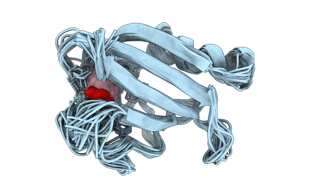
Title:
NMR structure of the ground state of the photoactive yellow protein lacking the N-terminal part
Biological Source:
Source Organism(s):
Halorhodospira halophila (Taxon ID: 1053)
Expression System(s):
Method Details:
Experimental Method:
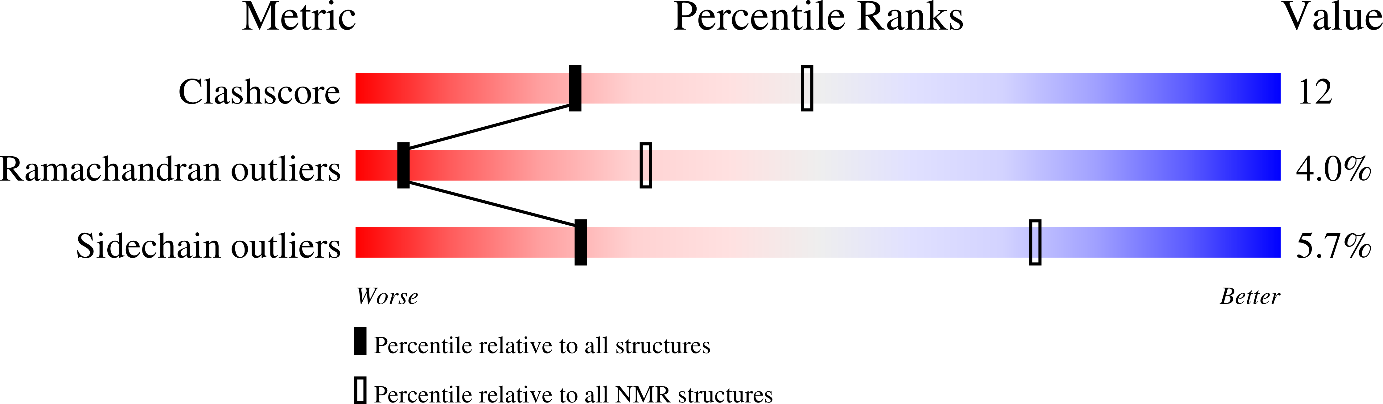
Conformers Calculated:
200
Conformers Submitted:
20
Selection Criteria:
structures with the least restraint violations