1XFG image
Deposition Date 2004-09-14
Release Date 2004-09-28
Last Version Date 2023-08-23
Entry Detail
PDB ID:
1XFG
Keywords:
Title:
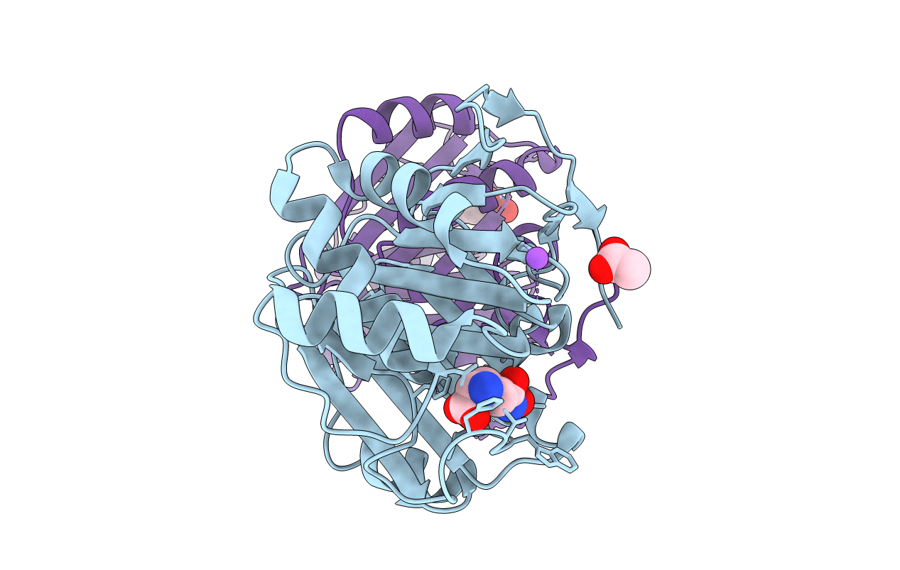
Glutaminase domain of glucosamine 6-phosphate synthase complexed with l-glu hydroxamate
Biological Source:
Source Organism:
Escherichia coli (Taxon ID: 562)
Host Organism:
Method Details:
Experimental Method:
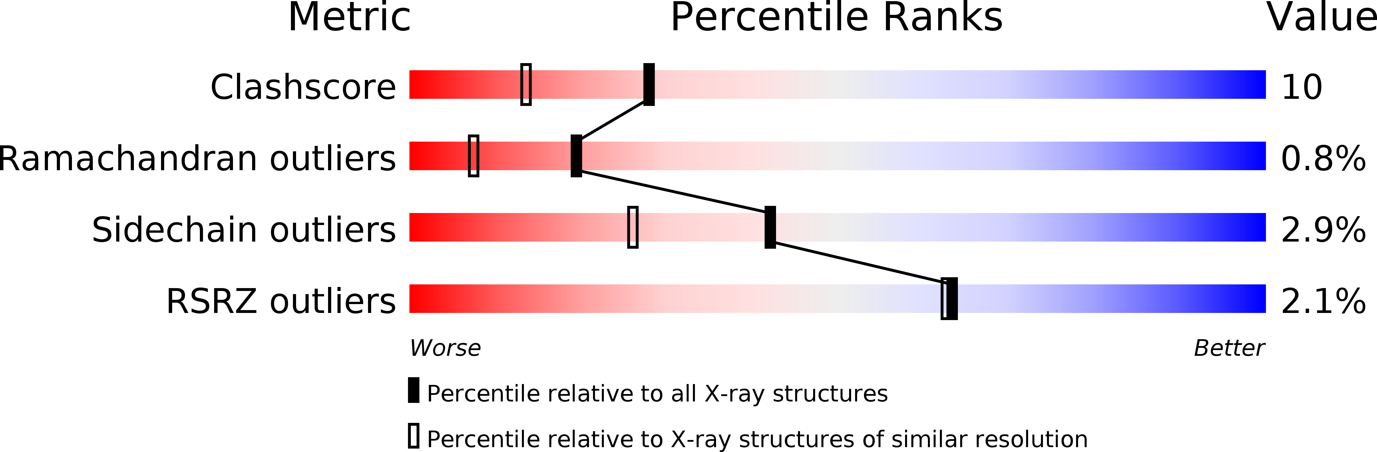
Resolution:
1.85 Å
R-Value Work:
0.15
R-Value Observed:
0.15
Space Group:
P 21 21 21
Macromolecular Entities
Structures with similar UniProt ID
Protein Blast
Polymer Type:polypeptide(L)
Molecule:Glucosamine--fructose-6-phosphate aminotransferase [isomerizing]
Gene (Uniprot):glmS
Chain IDs:A, B
Chain Length:240
Number of Molecules:2
Biological Source:Escherichia coli
Primary Citation
Substrate Binding is Required for Assembly of the Active Conformation of the Catalytic Site in Ntn Amidotransferases: Evidence from the 1.8 Angstrom Crystal Structure of the Glutaminase Domain of Glucosamine 6-Phosphate Synthase
Structure 4 801 810 (1996)
PMID: 8805567 DOI: 10.1016/S0969-2126(96)00087-1

Abstact

BACKGROUND Amidotransferases use the amide nitrogen of glutamine in a number of important biosynthetic reactions. They are composed of a glutaminase domain, which catalyzes the hydrolysis of glutamine to glutamate and ammonia, and a synthetase domain, catalyzing amination of the substrate. To gain insight into the mechanism of nitrogen transfer, we examined the structure of the glutaminase domain of glucosamine 6-phosphate synthase (GLMS). RESULTS The crystal structures of the enzyme complexed with glutamate and with a competitive inhibitor, Glu-hydroxamate, have been determined to 1.8 A resolution. The protein fold has structural homology to other members of the superfamily of N-terminal nucleophile (Ntn) hydrolases, being a sandwich of antiparallel beta sheets surrounded by two layers of alpha helices. CONCLUSIONS The structural homology between the glutaminase domain of GLMS and that of PRPP amidotransferase (the only other Ntn amidotransferase whose structure is known) indicates that they may have diverged from a common ancestor. Cys1 is the catalytic nucleophile in GLMS, and the nucleophilic character of its thiol group appears to be increased through general base activation by its own alpha-amino group. Cys1 can adopt two conformations, one active and one inactive; glutamine binding locks the residue in a predetermined conformation. We propose that when a nitrogen acceptor is present Cys1 is kept in the active conformation, explaining the phenomenon of substrate-induced activation of the enzyme, and that Arg26 is central in this coupling.

Legend

Protein

Chemical

Disease

Primary Citation of related structures