1XEG image
Deposition Date 2004-09-10
Release Date 2005-09-27
Last Version Date 2024-02-14
Entry Detail
PDB ID:
1XEG
Keywords:
Title:
Crystal structure of human carbonic anhydrase II complexed with an acetate ion
Biological Source:
Source Organism:
Homo sapiens (Taxon ID: 9606)
Method Details:
Experimental Method:
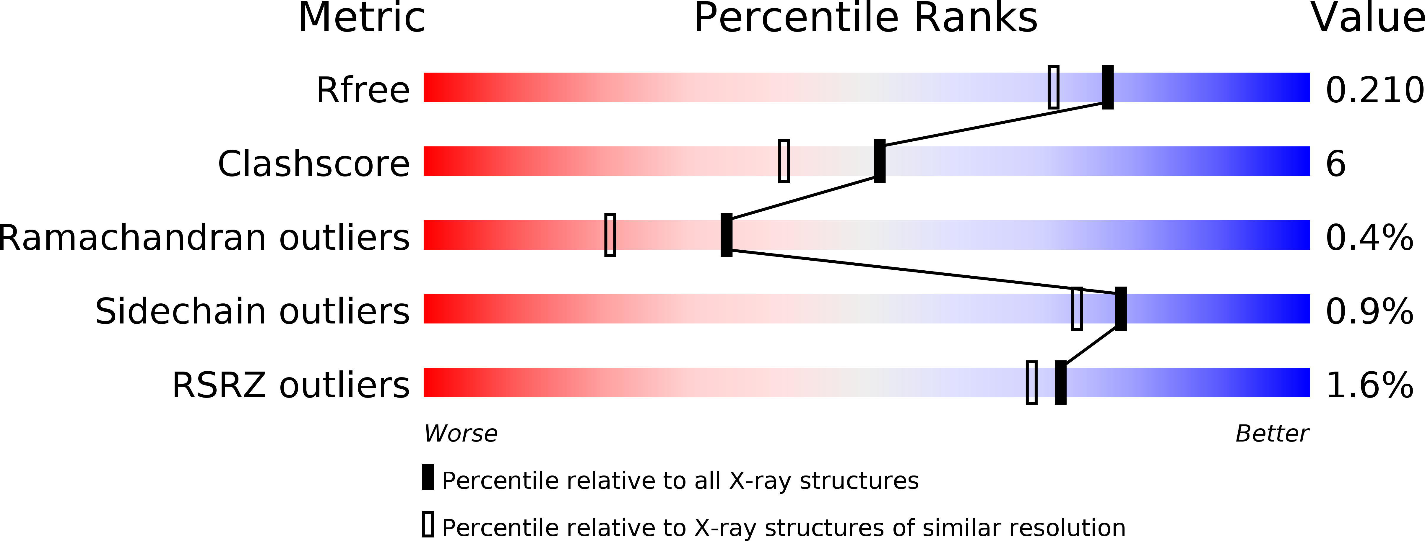
Resolution:
1.81 Å
R-Value Free:
0.21
R-Value Work:
0.18
Space Group:
P 21 21 21
Macromolecular Entities
Structures with similar UniProt ID
Protein Blast
Polymer Type:polypeptide(L)
Molecule:Carbonic anhydrase II
Gene (Uniprot):CA2
Chain IDs:A
Chain Length:260
Number of Molecules:1
Biological Source:Homo sapiens
Primary Citation
A novel acetate-bound complex of human carbonic anhydrase II.
Acta Crystallogr.,Sect.F 64 163 166 (2008)
PMID: 18323598 DOI: 10.1107/S1744309108002078

Abstact

The enzyme human carbonic anhydrase II (hCAII) crystallized in an acetate-bound complex belonging to space group P2(1)2(1)2(1), with unit-cell parameters a = 42.3, b = 71.8, c = 74.0 A. The structure was solved by the molecular-replacement method and refined to an R value of 0.18 and an R(free) of 0.21. The acetate molecule replaced the zinc-bound water molecule in the structure, differing from previous reports regarding the site of acetate binding. This mode of binding disrupts the hydrogen-bonded solvent network required for activity of the enzyme. This mode of inhibitor binding is a novel one that has not been observed previously.

Legend

Protein

Chemical

Disease

Primary Citation of related structures