
Deposition Date
2004-09-09
Release Date
2005-01-11
Last Version Date
2024-04-03
Entry Detail
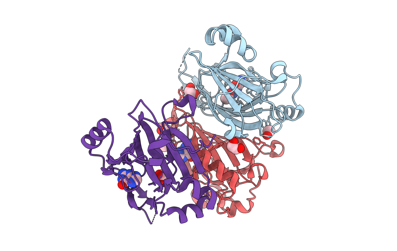
PDB ID:
1XE7
Keywords:
Title:
Crystal structure of the YML079w protein from Saccharomyces cerevisiae reveals a new sequence family of the jelly roll fold
Biological Source:
Source Organism:
Saccharomyces cerevisiae (Taxon ID: 4932)
Host Organism:
Method Details:
Experimental Method:
Resolution:
1.75 Å
R-Value Free:
0.24
R-Value Work:
0.22
R-Value Observed:
0.22
Space Group:
I 4 3 2
